임상 증상에 따라 서로 다른 치료 방법을 선택한 미로내 신경초종 2예
2 Cases of Intra-Labyrinthine Schwannomas With Different Treatment Options Based on Clinical Presentation
Article information
Trans Abstract
Vestibular schwannoma is a benign tumor that arises from the eighth cranial nerve and is the most common neoplasm in the internal auditory canal (IAC). It typically arises in the cerebellopontine angle or in IAC; however, in rare cases, they can arise within the labyrinth. These tumors arising in the labyrinth have been termed intra-labyrinthine schwannomas (ILS) and have different clinical features than the typical vestibular schwannomas. Clinical manifestations of an ILS typically include hearing loss, tinnitus, and vertigo, with unusual symptoms including a sense of fullness or imbalance. Temporal bone MRI is considered the most important test for diagnosing ILS. Tumors show nodular enhancement on T1-weighted images and filling defects on T2-weighted images. Treatment of ILS may include observation, microsurgical resection, and stereotactic radiosurgery depending on the location, size, and clinical presentation. In this paper, we present two cases diagnosed as ILS with different treatment options and provide a literature review for each case.
서 론
전정신경초종(vestibular schwannoma)은 제8 뇌신경에서 발생하는 느리게 성장하는 양성 종양이며 소뇌교각(cerebellopontine angele) 및 내이도(internal auditory cannal)에서 발생하는 가장 흔한 신생물이다. 그러나 이러한 전정신경 초종이 미로에서 발생하는 경우는 매우 드물다[1,2]. 이렇게 미로에서 발생한 전정신경초종을 미로내 신경초종(intralabyrinthin schwannoma)이라고 부르며, 일반적인 전정신경초종과는 구분되는 임상병리학적 특징을 가진 질환으로 인식되고 있다[3].
미로내 신경초종에 대하여 Kennedy 등[3]은 자기공명영상을 이용하여 7가지로 분류하였다. 전정내 신경초종, 와우내 신경초종, 전정와우내 신경초종, 와우축에 접한(transmodiolar) 신경초종, 평형반에 접한(transmacular) 신경초종, 골성이낭에 접한(transotic) 신경초종, 중이를 침범한(tympanolabyrinthine) 신경초종으로 분류하였다. 이 중 본 논문의 2예의 증례와 같은 전정내 신경초종이 전체 사례군 중 38%를 차지하여 가장 많다고 알려져 있다.
미로내 신경초종의 치료는 전정신경초종과 동일하게 위치, 크기, 그리고 임상 양상에 따라 경과 관찰(wait and scan), 미세수술적 절제, 또는 정위적 방사선 수술(stereotactic radiosurgery) 등을 고려할 수 있다. 그러나 유병율이 매우 드물기 때문에 아직까지는 치료방법을 결정하기 위한 요인이 불분명하다. 저자들은 미로내 신경초종으로 진단되었으나 임상증상에 따라 서로 다른 치료적 선택을 한 2예를 경험하였기에 이를 보고하는 바이다.
증 례
증례 1
70세 여자 환자가 1년 전부터 좌측 귀의 지속적인 이명 증상 및 어지럼증 증상이 발생하였으며 보행 시 보행장애 증상 호소하며 외래 내원하였다. 내원 당시 시행한 이학적 검사상 양측 고막 정상 소견이었으며, 청력검사에서 순음평균역치는 우측 25 dB, 좌측 72.5 dB을 보였다. 최대 어음 명료도는 우측 100%, 좌측 60%, 고막 임피던스 검사는 양측 정상 소견이었다. 온도안진 검사에서 좌측 반고리관은 우측에 비해 25% 정도의 마비 소견을 보였으며, 자발안진 및 체위 변화에 따른 안진은 보이지 않았다. 비디오 두부충동검사에서 양측 모두에서 시선재고정 단속운동(catch up saccade)은 관찰되지 않았으며, 전정유발 근전위 검사에서 좌측이 우측에 비해 63% 정도 진폭이 감소된 소견이 관찰되었다(Fig. 1). 측두골 자기공명영상검사(temporal bone MRI)를 시행하였는데 좌측 전정 내에 T1 강조영상에서 고신호강도로 관찰되며 T2 강조영상에서는 충만 결손 소견이 관찰되는 0.4 cm 정도 크기의 미로내 종괴가 발견되었다(Fig. 2).
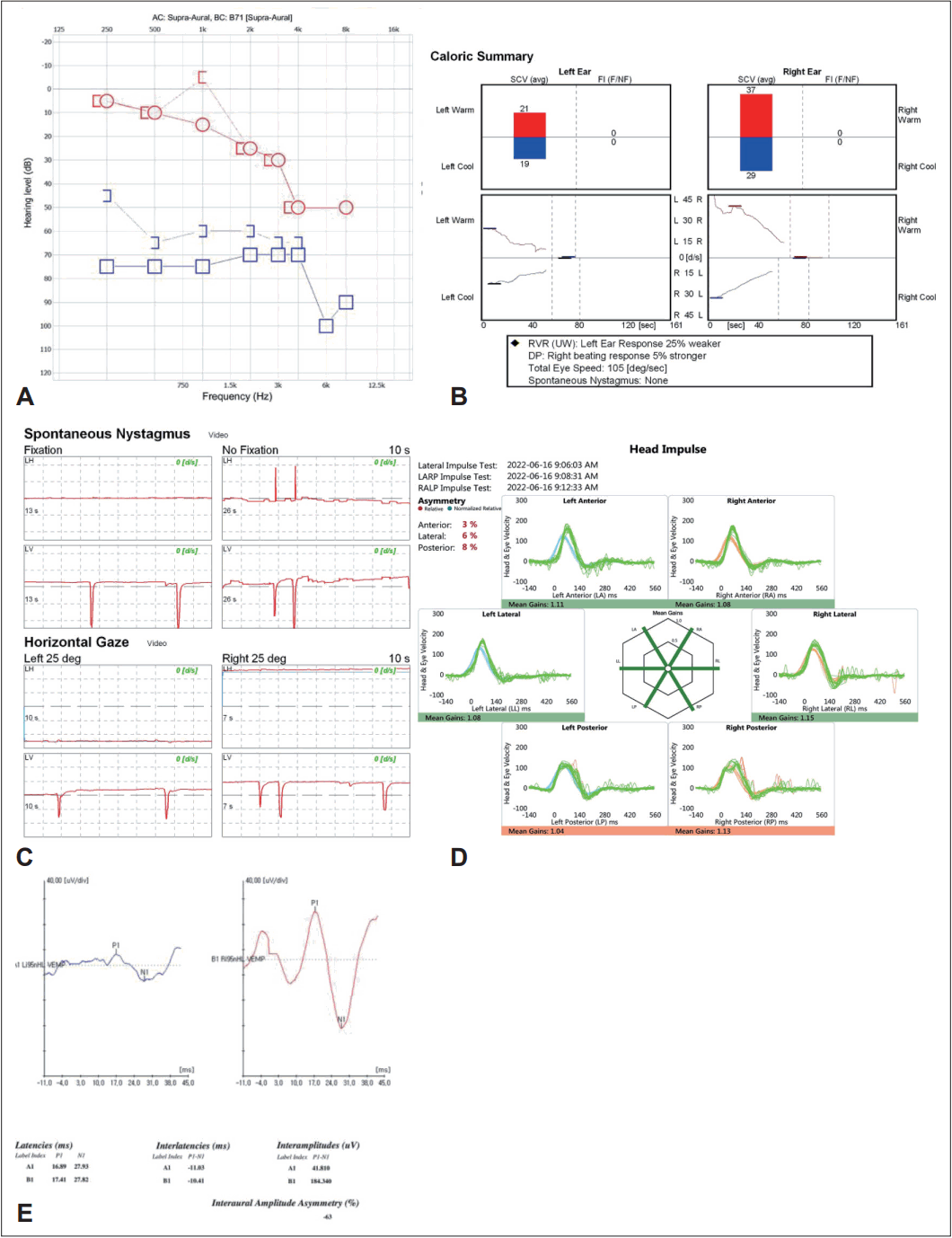
ENT examination performed on a patient with left intravestibular schwannoma. A: Pure tone audiometry. B: Caloric test. C: Electronystagmography. D: Head-impulse test. E: C-Vemp test.
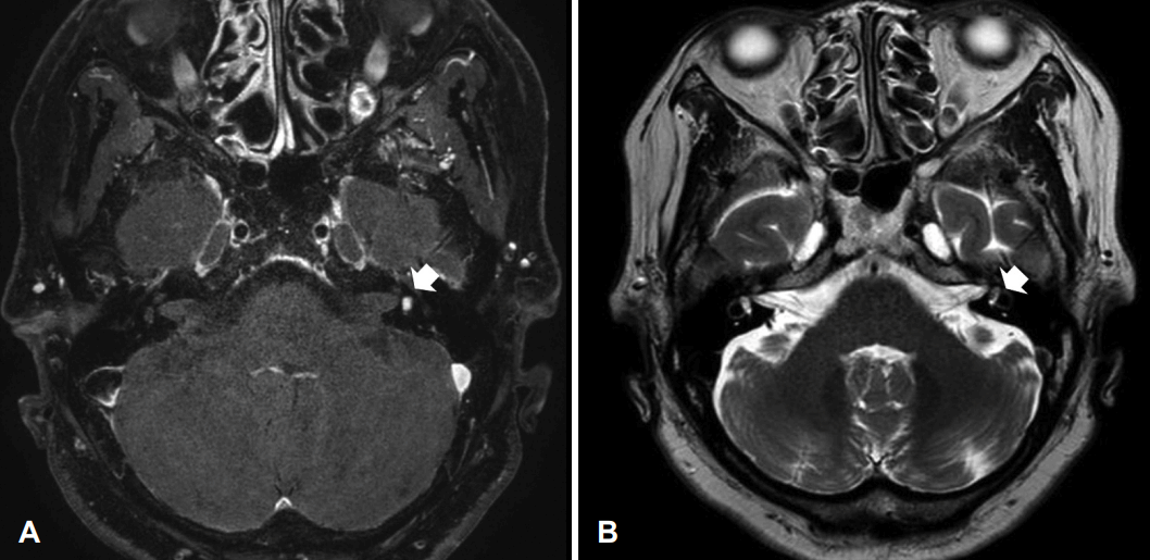
MR images. A: T1-weighted, Gd-enhanced axial image shows 0.4 cm sized focal enhancing mass in left vestibule (arrow). B: T2-weighted axial image shows a space-occupying lesion in the left vestibule (arrow).
이 환자는 전정 기능 저하와 난청이 심하지 않으므로 경과 관찰 하기로 결정하였다. 그러나 이후 5개월간의 추적 기간 동안 환자는 일상생활이 어려울 정도의 현훈 호소하며 보행 시 수차례 낙상을 경험하였을 뿐만 아니라 수술적 치료에 대한 강한 의지를 보여 수술적 치료를 결정하였다.
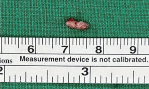
수술은 전신마취하에 진행하였으며 경유양동 접근을 통한 미로절제술(labyrinthectomy via transmastoid approach)을 시행한 후 결손부위는 서혜부 지방조직을 이용하여 보강하는 수술을 시행하였다. 반고리관을 노출시켜 공통각(common crus)과 전정(vestibule)을 노출시킨 후 전정으로부터 종물을 완전 절제 시행하였다(Fig. 3). 종물은 약 0.5 cm 정도 크기의 백색 양상이 확인되었다(Fig. 4). 수술 후 조직 검사에서 S-100 단백 염색에 양성반응을 보였고 Antoni type A와 B가 산재해 있어 신경초종임을 확인하였다(Fig. 5). 환자는 술후 7일째 특이소견 없이 퇴원하였다. 현재 6개월간의 추적 관찰 중에 있다.
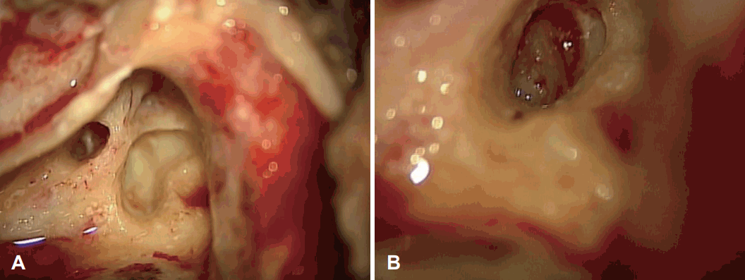
Intraoperative photographs after total labyrinthectomy. A: about 0.4 cm sized pin- kish mass filling the vestibule. B: The vestibule is exposed after the mass was completely removed.
증례 2
45세 여자 환자가 우측 귀 이명, 이충만감, 청력 저하 증상 발생하여 외래 방문하였다. 초진 때 시행한 신체검사에서 양측 고막은 정상 소견, 순음청력검사에서 우측 농, 좌측 8 dB을 보였다. 최대어음명료도에서 우측은 0%, 좌측은 100%의 소견을 보였고 고막 임피던스 검사상에서 양측 정상이었다. 비디오 두부충동검사에서 우측이 좌측에 비해 기능이 떨어진 것이 확인되었으며 시선재고정 단속운동관찰되었다. 전정 유발 근전위 검사에서 우측이 58% 정도 진폭이 감소된 소견이 관찰되었다(Fig. 6). 측두골 자기공명영상 검사에서 우측 전정 내에 T1 강조영상에서 고신호강도로 관찰되며 T2 강조 영상에서는 충만 결손 소견이 관찰되는 0.5×0.3 cm 정도 크기의 미로내 종괴가 관찰되었다(Fig. 7).

ENT examination performed on a patient with right intravestibular schwannoma. A: Pure tone audiometry. B: Head-impulse Test. C: C-Vemp Test.
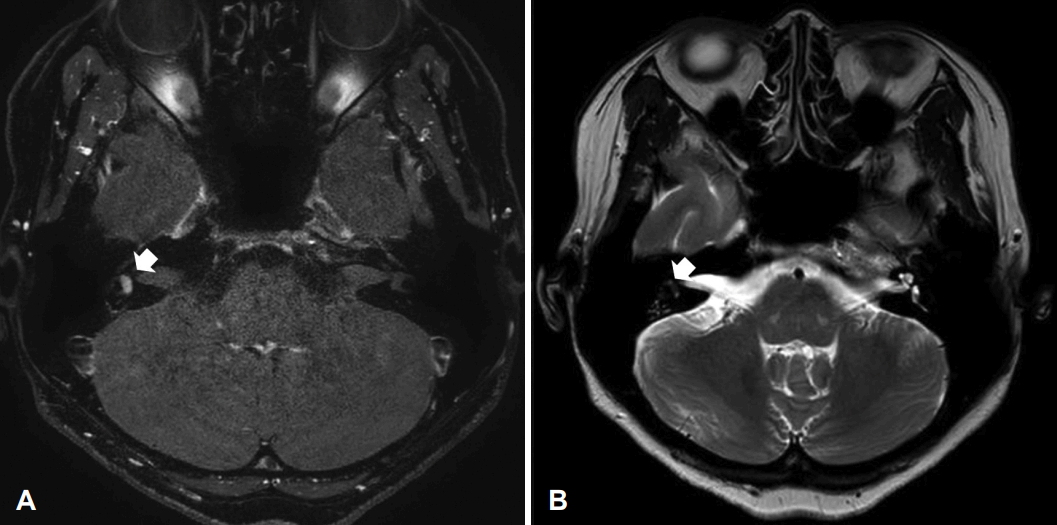
MR images. A: T1-weighted, Gd-enhanced axial image shows 0.5×0.3 cm sized focal enhancing mass in right vestibule (arrow). B: T2-weighted axial image shows a space-occupying lesion in the right vestibule (arrow).
이 환자의 경우 우측 심도난청과 전정 기능 저하 확인되어 수술적 치료를 고려하였으나, 평소 유발되는 어지럼증 없어 일상 생활에 있어 불편감이 없었을 뿐만 아니라 환자 침습적 수술에 대한 거부감 있어 경과 관찰을 결정하였다. 환자 6개월의 추적 관찰 기간 동안 병의 진행 없이 유지되고 있다.
고 찰
원발성 미로내 신경초종은 크기가 작고 서서히 자라는 경막외 종양으로 두개내에서 발생하는 종양에 비해 위험성이 상대적으로 낮다. 그러나 진행성의 청력소실, 이명, 현기증 및 이충만감 등의 증상을 유발하는 경우가 많으며, 이러한 임상 증상 및 작은 크기로 인해 미로내 신경초종은 메니에르, 전정편두통, 바이러스성 미로염 등 다른 질환으로 오인되는 경우가 많다[4].
미로내 신경초종에 대한 진단을 위해서는 조영제를 이용한 측두골 자기공명영상이 필요하다[5]. 종양은 일반적으로 T1 강조영상에서는 조영 증강을 보이며 T2 강조영상에서는 음영 결손을 보이는 양상을 보인다[6].
일반적으로 내이도 전정신경초종의 경우 수술적 치료시 청력을 보존할 수 없기 때문에 수술보다는 질병의 과도한 이환을 피하기 위해 MRI를 통해 정기적으로 관찰하는 경과 관찰을 선택하는 경우가 많다[6]. 그러나 조절이 되지 않는 난치성의 증상이 있을 경우 수술적 치료나 감마나이프 등의 적극적 치료를 고려할 수 있다[7,8]. 미로내 전정신경초종 역시 증상의 중증도와 종양의 위치에 따라 그 치료가 결정될 수 있으며 난치성 어지럼증이 있는 경우, 내이도 혹은 중이 내로 종양이 성장할 경우, 그리고 진단이 불확실할 경우 수술적 치료를 고려할 수 있다고 알려져 있다[9].
미로내 신경초종에서 난치성 어지럼증이 발생하는 기전은 아직까지 정확히 밝혀져 있지 않으나 몇 가지 가설이 존재한다. 종양의 성장에 따른 내이구조물의 변화나 전정신경의 압박이 중추신경계로 부적절한 신호를 전달하여 발생한다는 가설, 종양이 내이 임파액의 역동학을 변화시켜 세반고리관의 팽대부릉이나, 황반의 유모세포에 영향을 주어 발생할 것이라는 가설, 종양의 지속적인 염증이나 자극으로 인해 발생하였을 것이라는 가설 등이 제시되었다[10].
이러한 난치성 어지럼증이 동반된 미로내 신경초종에 대한 치료 옵션으로 생각해볼 수 있는 것은 외과적 제거, 감마나이프 등의 정위적 방사선 치료, 화학적 미로 절제술이 있다. 그러나 미로내 신경초종에 대한 정위적 방사선 치료에 대하여서는 사례가 극히 드물기 때문에 그 치료효과에 대하여 알기 힘든 상황으로 치료 옵션으로 선택하기 힘든 상황이다. 화학적 미로 절제술 역시 전정기능을 선택적으로 파괴하고 가능한 오랫동안 청력을 유지하기 위한 노력의 일환으로 고려 가능하나, 이러한 화학적 미로 절제술의 경우 증상의 개선시킬 수 있으나 종양의 크기를 조절할 수는 없다는 단점이 있다[4].
증례 1의 경우 종양의 크기는 0.5 cm 정도로 전정 내부에 국한된 종양이며 청력 역시 72.5 dB로 잔류 청력이 존재하여 경과 관찰을 고려하였으나, 난치성 어지럼증으로 인해 수차례 낙상을 경험하였으며, 환자가 수술적 치료를 통한 증상 완화에 대한 강한 의지를 가지고 있었기 때문에 난치성 현훈을 적응증으로 수술적 치료를 진행하였다. 이와 달리 증례 2의 경우 환자 우측귀 전농 및 전정기능 저하 소견으로 수술적 치료를 고려해볼 수 있었다. 그러나 종양의 크기가 크지 않고 현훈 등의 증상 역시 난치성으로 보이지 않았을 뿐만 아니라, 환자가 수술에 대하여 거부감이 있었다는 점 등을 고려하여 경과 관찰을 선택하였다.
미로내 신경초종은 미세한 크기로 인하여 진단이 어려우며 다른 질환과 비슷한 임상양상을 보인다는 점 때문에 MRI 기술이 발달하기 이전에는 진단이 어려웠던 질환이다. 최근 영상검사의 발달로 인해 미로내 신경초종의 진단은 점차 증가하고 있으며 앞으로도 계속 증가할 것으로 예상되고 있다. 이에 반해 아직까지 미로내 신경초종에 대한 치료적 결정을 하기 위한 명확한 가이드라인이 없는 상황이다. 이러한 상황 속에서 유사한 두 개의 미로내 신경초종 케이스에서 임상 증상에 따라 다른 치료적 결정을 적용하였기에 향후 미로내 전정신경초종의 치료가이드라인 도출에 있어서 도움이 될 수 있을 것으로 생각되어 보고하는 바이다.
Acknowledgements
None
Notes
Author contributions
Conceptualization: Jae Yong Byun. Data curation: Jong Hwan Lee. Ki-Jin Kwon. Formal analysis: Jong Hwan Lee. Methodology: Jae Yong Byun. Supervision: Jae Yong Byun. Writing—original draft: Jong Hwan Lee. Writing—review & editing: Jae Yong Byun, Jong Hwan Lee.

