이관 기능 검사
Eustachian Tube Function Test
Article information
Trans Abstract
Eustachian tube dysfunction (ETD) is a common condition in clinical otolaryngology practice. The eustachian tube (ET) is the only pathway connecting the middle ear cavity and the nasopharynx, and has a ventilation function to equalize the pressure of the middle ear and the atmosphere, a discharge function of middle ear secretions into the nasopharynx, and a defense function to prevent reflux from the nasopharynx. The most important function of the ET is to maintain a consistent middle ear air pressure for efficient sound transmission. Various methods have been used to measure the physiological function of ET. Traditional methods include the Valsalva, Toynbee, and Politzer test. Physiological methods include the 9-step Bluestone inflation-deflation test and sonotubometry. Evaluation of ETD using questionnaires is also being conducted, and currently, imaging tests are also conducted to evaluate the structure of ET. However, no standard protocols have been established until now. In this article, the types, methods, and advantages and disadvantages of the currently available ET function tests are introduced.
서 론
이관은 상기도의 확장된 구조물로 중이와 비인강을 연결하는 유일한 통로이다[1]. 이관은 좁고 긴 관의 형태로 외측 1/3의 골부와 내측 2/3의 연골부로 형성되어 있으며, 길이는 31-38 mm이다[2]. 정상적인 이관 구조에서 중이강 측의 골부는 항상 열려 있으나, 인두편도의 외측에 존재하는 비인강측의 연골부는 평상시에는 주로 닫혀 있다[3]. 이관의 중요한 기능은 다음과 같다. 첫째는 중이의 환기(ventilation) 기능이다. 이관은 평상시에는 폐쇄되어 있으나, 하품, 연하, 재채기 등 후에는 연골부 이관이 일시적으로 열리면서 중이 내 압력과 대기압의 균형을 유지하게 된다. 또한 중이강 내의 공기는 중이 점막을 통한 확산(diffusion)에 의해 흡수되어 점차적으로 음압으로 변하게 되는데, 이관 개방을 통한 신선한 공기를 주입함으로써 일정한 중이압(-50~50 mm H2O)을 유지하는 데 도움을 준다[4,5]. 두 번째는 중이의 정화(clearance)이다. 중이강에서 비인강까지 분포되어 있는 점액막층(mucous blanket)의 점액섬모(mucociliary)가 능동적으로 이물질을 비인강쪽으로 분비하는 역할을 한다. 마지막은 중이의 방어 역할이다. 평상시 비인강 측의 이관 폐쇄로 비인강에서 발생하는 소리 혹은 이물질의 역류로부터 중이를 보호해준다[6,7]. 이렇듯 다양한 이관 기능이 정상적으로 유지되어야 염증에 대한 방어가 가능하며, 중이의 압력을 일정하게 유지함으로써 효과적으로 소리를 전달할 수 있다. 이관이 잘 열리거나 닫히지 않는 기능부전이 발생하면 귀먹먹함이나 자가강청 등 다양한 증상이 발생할 수 있으며, 최근에는 이러한 증상에 대한 관심이 점차 높아지면서 이관풍선확장술이나 이관 폐쇄술 등 적극적인 수술적 치료가 많이 이루어 지고 있다[8,9]. 그러나 아직까지도 이관의 기능을 평가하는 방법은 정립되어 있지 않다. 따라서 본 종설은 이관기능부전을 평가할 수 있는 유용한 방법에 대해 고찰해 보고자 한다.
이관 통기 검사
Valsalva 검사
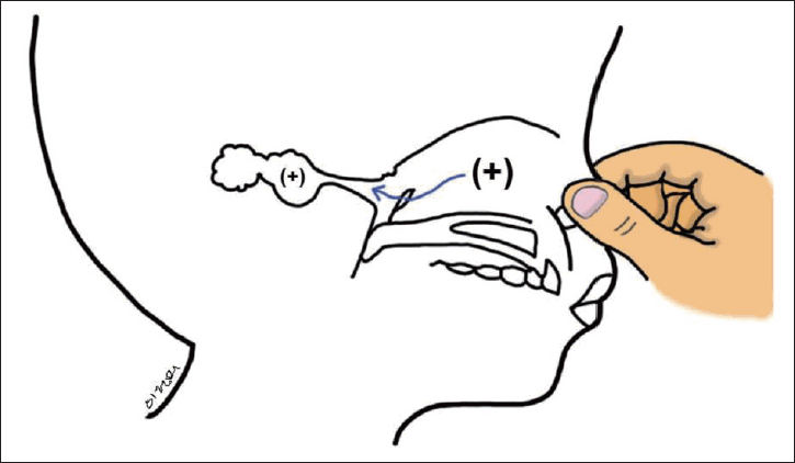
Valsalava 검사는 강제 호기(입을 닫고 엄지와 검지로 코를 잡은 상태)에 의해 이관과 중이가 팽창될 수 있는 경우 양성(정상)으로 간주한다(Fig. 1). 이렇게 생성된 고압은 가변적이며, 최대 2000 mm H2O이다.

Valsalva test. The lumen of eustachian tube is opened by a forced expiration with the nose closed by finger pressure and the mouth closed, which insufflates positive pressure into the middle ear through the eustachian tube.
외이도에 양압이 가해지면 고막이 안쪽으로 움직이지만, 중이에 양압이 가해지거나 외이도에 음압이 가해지면 고막이 바깥쪽으로 움직인다. 고막이 손상되지 않은 경우, pneumatic otoscope을 사용하여 중이에 양압이 가해질 때 고막이 부풀어 오르는 것을 관찰할 수 있다[10].
중이 압력의 변화는 압력계(manometry) 또는 고실계측법으로 확인할 수 있다. Valsalva 검사 시 중이에 생성되는 양압은 일시적으로, 대부분의 정상적인 이관 상태에서는 이관이 팽창한 후 닫히기 전에 즉각적인 압력의 평형이 이루어진다. 따라서, 중이 압력의 변화는 고실계측법에 의해 기록되지 않을 수 있다. 고막이 손상되지 않은 상태에서 중이로 들어가는 공기 소리를 청진기나 토인비관으로 들을 수 있으나, 이러한 방법은 더 이상 잘 사용되지 않는다.
Valsalva 검사 결과는 이관 압력 조절 기능을 판단할 수 있는 방법은 아니다. Valsalva 검사 결과가 양성이면 해부학적으로 정상적인 이관 연결 통로가 있고, 비인강 압력 변화 시 확장될 수 있는 이관 상태임을 암시한다. 실제로, 검사하는 동안 중이의 팽창 없이는 이관 기능에 관한 유용한 정보를 얻을 수 없다. Elner 등[11]은 정상적인 귀를 가진 101명의 성인 중 85%가 Valsalva 검사 양성이 나타났음을 보고 했다. Bunne 등[12]은 정상적인 고막 소견을 보이는 그룹에서 Valsalva 검사 시 중이염 및 고막 함몰이 있는 그룹보다 유의하게 양성 반응이 높게 나왔음을 보고했다. Canali 등[13]은 정상적인 고막 상태에서 Valsalva 검사 반응이 잘 나타났으며, 고막 함몰 및 변형이 심해질수록 검사 양성 반응이 떨어지는 것으로 보고했다[13].
Toynbee 검사
대상자는 코를 막은 상태에서 침을 삼키도록 하여 비인두 내에 양압을 생성한 다음 음압으로 만든다(Fig. 2) [14]. 중이 압력의 변화는 Valsalva 검사에서 평가되는 것과 동일한 방식으로 평가한다. 중이 내 압력이 음압이 되는 경우, 고막은 수축되고 pneumatic otoscope으로 외이도에 양압을 가했을 때 고막은 안쪽으로 이동하지 않는다. 만약 중이 내에 형성된 음압보다 더 낮은 압력을 외이도에 가하면 고막은 바깥쪽으로 이동한다. 고막이 천공되어 있어도 중이강 내의 압력의 변화를 압력계로 측정하여 이관의 환기 기능을 확인할 수 있다.

Toynbee test. A: Positive pressure phase. Closed-nose swallowing results first in positive pressure in the nose and nasopharynx. B: Negative pressure phase. During swallowing, the positive pressure in the nasal cavity changes to negative pressure.
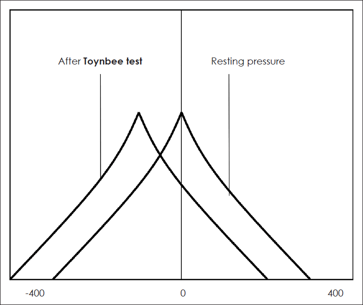
검사 결과는 일반적으로 중이 압력에 변화가 있을 때 양성으로 판단한다. Toynbee 검사 후 중이 내 음압 또는 정상 중이 압력 후에 나타나는 일시적인 중이 내 음압은 이관이 생리적으로 좋은 기능을 하고 있는 것을 의미한다(Fig. 3). 이는 비인강 압력 변화에 따라 이관이 활발하게 열릴 수 있다는 뜻이다. 하지만 Toynbee 검사를 시행하는 동안 중이 압력 변화가 없어도 이관 기능이 저하되었다고 판단할 수 없다. 과거 연구에서 이관 기능 저하로 판단되는 성인의 30%가 Toynbee 검사에서 정상을 보이는 것으로 보고했다[15]. Elner 등[11]은 Toynbee 검사가 정상 성인의 79%에서 양성이었다고 보고했다. Bluestone 등[16]은 외상성 천공이 있는 “정상” 어린이 7명 중 6명에서 Toynbee 검사 양성으로 나타났지만, 고막 함몰 또는 진주종이 있는 어린이에서는 Toynbee 검사 후 압력 변화가 없었다고 보고했다.

Tympanogram obtained before and after the Toynbee test. The negative middle-ear pressure is considered to be associated with good Eustachian tube regulation function.
Toynbee 검사는 소아보다 성인에서 이관 기능을 판단하는데 유용하다. 하지만 대상자의 연령에 관계없이 검사 중 또는 검사 후에 중이에 음압이 발생하면 이관이 활발하게 열리는 것으로 판단할 수 있다. 또한 비인강 음압을 견딜 수 있을 만큼 충분히 이관의 구조가 단단하다는 것(비인강 내 음압 변환시 이관이 완전히 닫히지 않음)을 의미하기 때문에 이관 기능이 정상적으로 작동하는 것으로 볼 수 있다. 하지만 중이 내 양압이 나타나거나 압력의 변화가 없다고 해도 이관 기능이 비정상이라고 단정할 수 없기 때문에 다른 이관 기능 검사를 시행할 필요가 있다.
Toynbee 검사 방법
1) 코를 막고 삼키면 코와 비인두에 양압이 발생한다(positive pressure phase).
2) 이후에 비인강의 음압으로 바뀐다(negative pressure phase).
3) 비인강 내 양압 상태일 때 공기가 중이로 들어가 양압을 생성한다.
4) 이후 비인강 내 음압 단계 동안 또는 이후에 중이에 음압이 발생할 수 있다. 또는 중이에 양압이 그대로 존재하거나, 양압 이후에 중이 내 음압이 뒤따를 수 있다.
5) 이관이 양압 및 음압 단계에서 열리지 않으면 중이 압력의 변화가 발생하지 않는다.
Politzer 검사
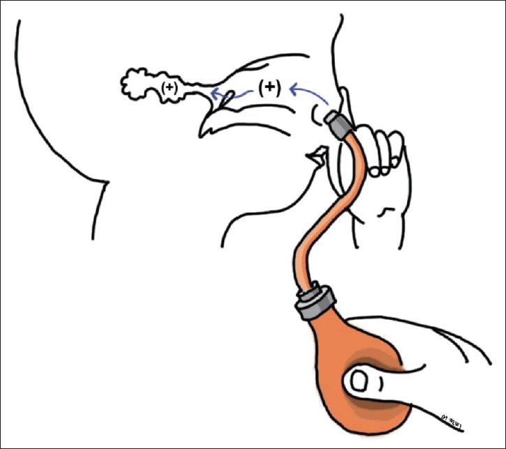
Politzer 검사는 공기 주입기가 달린 고무구를 한 쪽 콧구멍이 넣어 공기가 새어나가지 않도록 압박하면서 반대쪽 콧구멍을 손가락으로 누른다. 대상자에게 침을 삼키게 하거나, ‘ㄱ’과 같은 발음을 시키면 연구개가 상승되어 비인두를 폐쇄시킨다. 이 때, 고무구를 압축하여 비인강 내로 공기를 넣어주어 이관을 열리게 하는 방법이다(Fig. 4) [17]. 비인강에서 제공되는 양압이 중이로 전달되어 중이 내 압력이 상승하면 검사 양성으로 판단할 수 있다. 정상 검사 결과의 유의성은 이관의 환기 기능이 있다고 판단할 수 있는 점이지만 이관의 생리적 기능을 평가하지는 못한다.

Politzer test. The “Poltizer bag” is inserted into one naris while both nasal alae are compressed by finger pressure. The patient is asked to repeat the letter K or is asked to swallow, both of which close the velopharyngeal port, while the examiner compresses the “Poltizer bag.” When normal eustachian tube function is present, positive pressure is insufflated into the middle ear through the Eustachian tube.
고막운동성계측법(Tympanometry)
고막운동성계측법은 현재 폐쇄성 이관 기능 평가에 일차적으로 사용되는 검사이다. 중이 내 압력은 정상 성인에서 -50~50 mm Hg 사이로 나타난다. 고막운동성계측법은 중이 삼출물을 감지하는 데 효과적이며 보고된 민감도와 특이도는 각각 94%와 95%이다. 그러나 이관기능부전 시 측정된 중이강 내 압력은 외부 압력이 안정적인 상황에서 압력에서 보통 정상으로 나타난다[18]. 과거 연구에서 이관기능부전 군과 정상 군의 중이 내 압력을 측정했을 때 안정 상태에서 유의한 차이가 없는 것으로 보고했다[19,20]. 중이 압력의 일회성 측정으로 이관의 개방 및 기능에 대한 정보를 확인할 수 없다. 고막운동성계측법으로 측정한 중이 압력은 측정 시간마다 변동할 수 있기 때문에 변동되는 값을 통해 중이 내 압력 범위를 예측해야 한다[21].
9단계 고실계측도 검사
이관 기능을 측정하는 방법으로 Bluestone이 개발한 inflation-deflation test가 있다. 고막이 손상되지 않은 상태에서 이관 기능을 평가하는 데 유용하다. 중이 내 삼출액은 없어야 하며 9단계 고실 측정 절차는 다음과 같다(Fig. 5) [22,23].

Bluestone nine-step inflation-deflation tympanometric test. EAC, external auditory canal; ET, Eustachian tube; ME, middle ear; TM, tympanic membrane; TVP, tensor veli palatini muscle.
1) 안정 상태의 중이 압력 고실도를 확인한다.
2) 외이도에 200 mm H2O의 압력을 가해 고막을 내측으로 편향 시키면 중이 내 압력이 증가한다. 대상자는 중이 내의 높아진 압력을 낮추기 위해 침을 삼킨다.
3) 대상자가 침 삼키기를 멈추고 있는 동안 외이도에서 가해진 압력을 정상으로 되돌리면 고막은 외측으로 편향되며 중이 내 압력은 약간의 음압을 보이게 된다. 이 때 고실도는 중이 내 음압 상태로 표시된다.
4) 대상자는 중이 내 압력의 평형을 유지하기 위해 침을 삼킨다. 비인두에서 중이 내로 공기가 통하면서 중이 내 압력이 정상화된다.
5) 이 때 고실도는 정상 압력 상태로 나타난다.
6) 외이도에 -200 mm H2O의 압력을 가해 고막을 외측으로 편향시키면 중이 내 압력이 감소한다. 대상자가 중이 내 음압을 정상화하기 위해 침을 삼킨다. 비인두에서 중이 내로 공기가 통하면서 중이 내 압력이 정상화된다.
7) 외이도에서 가해진 음압을 정상으로 되돌릴 때, 대상자는 침 삼키기를 멈춘다. 이 때 고막이 내측으로 이동하면서 중이 공간은 양압 상태로 변하고, 고실도 또한 양압 상태로 표시된다.
8) 대상자는 중이 내 양압을 줄이기 위해 침을 삼킨다. 이때 중이에서 비인두로 공기가 통하면서 중이 내 압력이 정상화된다.
9) 최종적으로 고실도는 정상 압력 상태로 기록된다. 이 검사법은 이관기능장애를 평가할 수 있는 간단하고 유용한 방법이다. 환자가 nine-step test의 모든 단계를 통과한다면 이관 기능이 양호한 것으로 판단할 수 있다. 대부분의 정상적인 성인은 수행할 수 있으나, 아동의 경우에는 정확한 평가가 어려울 수 있다.
McBride 등[24]은 107명의 성인을 대상으로 두 가지의 비침습적인 방법으로 이관기능평가를 시행했다. Bluestone 9단계 inflation-deflation 검사와 sonotubometry를 사용하여 평가했는데, 두 가지 검사를 병합했을 때 96%의 정상 이관 기능을 확인할 수 있었다.
변형 가압-감압 검사
Inflation-deflation test는 Miller가 소개한 방법으로 현재 이를 바탕으로 변형된 검사를 시행하고 있어 modified inflation-deflation test라고 한다[25,26]. modified inflation-deflation test는 고막 천공 또는 고막 환기관 삽입이 되어있는 경우, 고실계측기의 압력계를 이용하여 이관 환기 기능을 측정하기 위해 시행할 수 있다. 외이도를 압력 측정 튜브가 달린 실리콘 귀마개로 밀폐하고 이 튜브를 통해서 중이강에 400~-200 mm H2O의 압력을 가하면서 이관의 압력 조절 기능을 평가한다[27,28].
중이에 양압이 가해질 때(inflation) 이관은 수동적 또는 능동적으로 열릴 수 있다. 이관이 강제로 열릴 수 있도록 중이에 충분한 양압이 가해져야 하는데, 이관이 수동적으로(passive opening) 열린 후 중이에 남아 있는 압력을 closing pressure라고 한다. 이후 침 삼키기(active opening)에 의해 tensor veli palatini muscle이 수축하면 이관의 내강이 열리면서 추가적인 압력 평형이 이루어진다. 중이 내 압력 변화 과정은 고실계측기로 압력을 측정할 수 있으며, 이관의 수동 및 능동적 열림 이후 중이에 남아 있는 압력을 residual positive pressure 라고 한다. 반대로 중이에 음압을 가한 후(deflation) 침 삼키기(active opening)를 통해 압력 조절을 유도하면서 변화 과정을 측정하고, 남아 있는 residual positive pressure 를 확인한다. Residual pressure가 고실계측도상 0 mm H2O에 근접하여 나타나면 압력 평형이 이루어진 것으로 판단할 수 있다(Fig. 6).

The results of modified inflation-deflation pressure regulation tests in normal adult with a traumatic perforation. C, closing pressure; O, opening pressure; RP, residual pressure; S, pressure after swallow.
Modified inflation-deflation test를 통해 이관의 생리학적 기능을 완벽히 판단할 수 없지만, 이관의 정상 또는 비정상 기능을 구분하는 데 도움이 된다. 검사 결과 정상 범위 내에서 passive opening과 closing pressure를 나타내고, 침 삼키기 이후 residual pressure가 평형을 이루며, 음압을 가한 후에도 평형상태로 회복될 수 있다면 정상 이관 기능으로 판단할 수 있다. 음압 상황에서 고려할 점은 침 삼키기 후에 중이 내 압력이 일부만 평형을 유지하거나, 평형으로 돌아오지 않을 수 있다. 이는 음압이 빠르게 가해질 때 정상적인 이관도 닫힐 수 있기 때문이다[29].
설문지를 이용한 평가
이관기능평가와 관련된 설문 조사는 7-item Eustachian Tube Dysfunction (ETD) Questionnaire (ETDQ-7) 및 10-item Cambridge ETD Assessment (CETDA)가 있다(Tables 1 and 2) [30,31]. ETDQ-7은 영어, 독일어, 네덜란드어, 덴마크어 및 포르투갈어로 검증되었으며, 다양한 임상 연구의 결과 척도로 사용되었다[32-34]. 최근 대만에서도 ETDQ-7 검증연구를 시행했으며, 이관기능저하의 증상의 중증도를 정량적으로 평가할 수 있는 신뢰성 있는 검사로 보고하였다[35].
McCoul 등[31]은 이관기능저하를 진단 받은 성인 50명과 대조군 25명을 대상으로 ETDQ-7을 처음과 4주 후에 반복 검사했다. 검사-재검사 신뢰도는 4주 간격으로 작성한 두 설문지에서 좋은 상관관계를 보였다(r=0.78). ETDQ-7은 이관기능저하가 환자를 구별할 수 있어(p<0.001) 진단에 유용한 것으로 보고했다. ETDQ-7의 정확도를 검증한 다른 연구에서 ETDQ-7의 민감도와 특이도는 각각 70% 및 100% 나타났다. 하지만 실제 이관기능장애를 진단할 수 있는 객관적인 척도는 떨어지며, 이관기능장애의 증상을 잘 반영하는 것으로 보고했다[32].
10개 항목으로 구성된 CETDA는 보다 최근에 개발 및 검증되었다[31]. CETDA는 일관성 및 재현성 면에서 ETDQ-7과 비슷한 효과를 보이는 것으로 나타났으며, 두 가지 방법으로 측정된 점수는 상관성이 있었다. 그러나 두 가지 검사법을 통해 폐쇄성 이관기능저하와 이관개방증을 감별하기는 어려운 것으로 보고되었다[36,37].
하지만 이관풍선확장술 전후에 시행한 ETDQ-7은 이관기능저하 증상의 호전에 따라 차이가 나타났다[38,39]. 두가지 설문지 평가는 ETD 관련 증상 및 중증도를 정량화하는 데 유용하다. 따라서 설문 조사 평가는 진단적 유용성은 다소 떨어지나 이관 기능에 대한 다양한 시술 후 ETD 증상의 변화를 평가할 때 사용할 수 있겠다[40].
음향이관측정법
이관을 통한 소리의 전도는 Politzer에 의해 처음 발견되었다. 코 근처에서 발생한 소리굽쇠의 소리가 삼키는 동안 진폭이 증가하는 것을 관찰했다. Politzer는 소리가 침을 삼키는 동안 열리는 이관을 통해 이동하면서 발생한 현상으로 생각했다. 1932년, Gyergyay는 다양한 악기를 사용하여 코와 이관을 통해 소리가 전달되는 것을 보고하였다[41].
Virtanen은 1977년에 광범위한 주파수 대역을 사용하여 이관 기능을 평가했다. 1-20 kHz의 single tone을 1 kHz 간격으로 제공했을 때 이관을 통한 소리 전도는 6, 7, 8 kHz에서 가장 잘 나타났다. 또한 삼킴으로 인한 생리학적 소음을 기록했으며 최대 5 kHz까지 나타났다. 따라서, 5 kHz 미만의 주파수를 사용한 소리 전도는 삼킴으로 인한 생리학적 소음에 의해 왜곡되기 때문에 유효하지 않다고 보고했다[24].
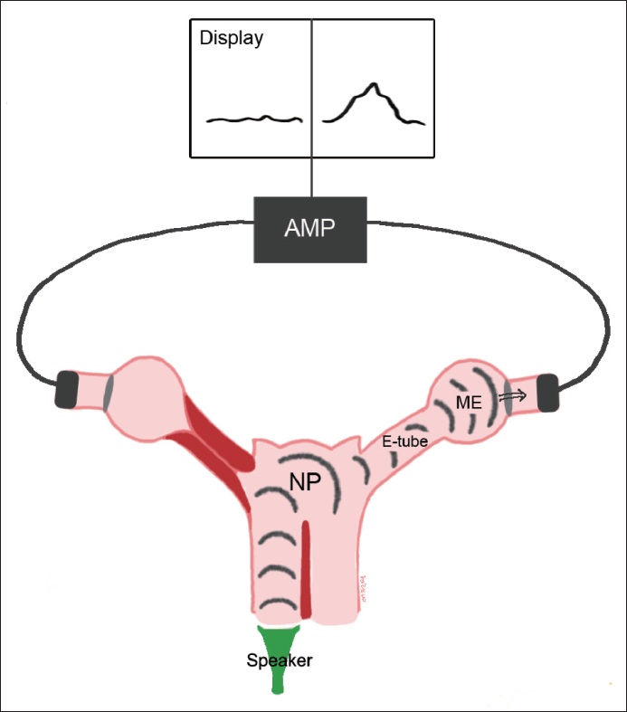
Sonotubometry는 이관을 통한 소리의 통과 정도에 따라 이관 기능을 판단한다. Tone generator에 연결된 소형 휴대용 스피커에서 발생하는 소리를 이용하여 검사를 진행한다. 코의 입구에서 3~10 kHz 순음 또는 백색 잡음(white noise)을 제공한다. 소리는 이관이 열릴 때 코와 이관을 거쳐 외이도로 전달된다. 양측 외이도에 배치된 microphone을 통해 신호를 수신하고 증폭하여 기계에 표시한다. Sonotubometry는 이관 개방으로 인한 외이도의 소리 증가를 감지하고 디지털화하여 저장한다. 저장된 정보에는 전송된 소리와 시간의 따른 음량 변화가 기록된다. 표현된 그래프는 이관이 열리는 시간과 정도에 대한 정보가 포함된다(Fig. 7).

A schematic diagram of sonotubometry. AMP, amplifier; NP, nasopharynx; E-tube, Eustachian tube; ME, middle ear.
Sonotubometry의 장점은 이관의 환기 기능을 비침습적으로 측정 가능하다는 것이다. 이관 기능 측정은 침 삼키기와 같은 생리적인 상황에서 이루어지며, 압력 자극이 필요하지 않다. 또한, 고막 천공이 있는 경우에도 측정이 가능하다. Jonathan 등[42]은 건강한 성인에서 이관 기능을 평가하기 위해 음파 측정과 고실계측법을 비교했으며, 두 방법 간에 높은 상관관계를 보이는 것으로 보고했다. 이후 Jonathan [43]은 소아를 대상으로 건강한 군과 중이 삼출액이 있는 군에 대해 이관 기능을 평가하였다. Sonotubometry와 고실계측법, 청력검사를 시행했을 때, 모든 소아에서 sonotubometry가 가능했음을 보고했다.
결론적으로, sonotubometry는 고막 손상 유무에 관계 없이 이관 기능 평가를 할 수 있다. 또한, 통증 없이 검사를 시행할 수 있어 성인과 어린이 모두에게서 평가하기 쉽다. Sonotubometry의 재현성은 다른 이관 기능 검사만큼 높은 것으로 나타났다. 하지만 중이압을 유지하기 위해 24시간 동안 이관이 얼마나 열려야 하는지는 정확히 알려지지 않았기 때문에, sonotubometry의 결과를 평가할 때 침 삼키기 후에 이관이 항상 열리지 않는다는 점을 고려해야 한다.
Valsalva를 동반한 측두골 컴퓨터단층촬영
이관 기능 평가 및 시각화를 위해 측두골 컴퓨터단층촬영(CT) 시 환자에게 valsalva 동시에 시행하는 검사가 시행되고 있다. 다중 평면 재구성을 포함하는 고해상도 CT가 개발되면서 이관의 3D 해부학 및 주변 조직, 길이와 방향 평가에 대한 연구가 점차 확산되었다[44,45]. Valsalva 시행 시 생리학적으로 협착되어 있는 이관의 말단 부분 연골부를 시각화할 수 있는 장점이 있다. 먼저 정상 조건에서 측두골 CT를 시행한 다음 환자가 Valsalva를 수행하는 상태에서 촬영을 다시한다. 두 번째 검사 시 코를 닫은 상태에서 강하게 내쉴 수 있도록 교육이 필요하다[46,47].
Tarabichiet와 Najmi [47]는 이관 내 연골부를 시각화하기 위해 Valsalva 기법과 함께 측두골 CT의 유용성을 평가했다. Valsalva 시행 후 검사된 귀의 27/76(35%)에서 이관 전체 길이를 시각화할 수 있었다. 71/76(94%) 귀에서 이관 연골부의 1/3을 시각화할 수 있었다.
또다른 연구에서 이관기능설문조사와 Valsalva 측두골 CT의 연관성을 분석하였다. Valsalva 시행 후 ETDQ<7 군에서는 33%의 환자에서 이관을 영상으로 확인할 수 있었다. ETDQ≥7 그룹에서는 Valsalva 시행 후 29%에서 이관을 확인할 수 있었다. 두 그룹 간에 Valsalva 시행 전후의 이관의 시각화는 유의한 차이가 없었다[46].
Kourtidis 등[48]은 일측성 이관기능부전 환자에 대해 측두골 CT 및 Valsalva를 시행한 전향적 연구를 시행했다. 결과적으로 영상학적 평가의 민감도와 특이도가 각각 52.5%와 97.5%로 임상에서 이관기능장애에 대한 선별 검사로는 부적절하다고 보고하였다.
최근 발표된 연구에서 Valsalva-측두골 CT로 얻은 이관의 모양과 ETD 점수와의 상관관계를 분석하였다. 고해상도 영상학적 검사로 이관의 복잡한 해부학적 구조에 대해 자세한 정보를 얻을 수 있었다. 그러나 측정된 이관의 형태와 설문지 점수 사이에 유의한 상관관계가 나타나지 않았다. 따라서 이관의 3D 영상 측정으로 이관기능을 평가하기에는 부적절한 것으로 보고하였다[49].
결 론
임상 현장에서 이관기능저하를 호소하는 환자는 언제든지 마주칠 수 있다. 최근에는 이관풍선확장술과 같은 수술법이 신의료기술로 인정되면서 정확한 이관기능평가에 대한 관심이 더욱 높아지고 있다. 이관을 통한 압력 조절 기능은 다양한 방법을 통해 평가가 이루어졌다. 하지만 현재까지 알려진 이관기능검사 중 한 가지 방법만으로 이관기능을 정확히 평가할 수는 없다. 다양한 임상 연구 결과, 두 가지 이상의 검사법을 함께 적용하는 것이 이관기능평가의 정확도를 높이는데 도움이 되는 것으로 밝혀졌다. 중이검사기계를 이용하여 9 step inflation-deflation 검사와 환자 설문 조사 조합이 높은 이관기능저하 진단률을 보이는 것으로 알려져 있다. 이관 기능저하에 대한 설문지 검사는 다수의 외국에서 번역 및 검증을 통해 이용하고 있다. 국내에서도 이관기능저하 설문지 검사에 대한 번역 및 검증이 이루어진다면 임상 현장에서 유용하게 이용될 수 있을 것이다. 또한, sonotubometry와 같이 비침습적인 평가 기기가 도입된다면 이관기능검사에 더욱 쉽게 접근이 가능할 것이다. 현재까지 알려진 이관기능평가 검사법에 대해 정확히 이해하고, 새롭게 개발되는 검사법에 대한 연구가 활발히 이루어진다면 이관기능저하에 대한 이해와 적절한 치료가 가능할 것으로 기대된다.
Acknowledgements
This work was supported by a National Research Foundation (NRF) of Korea grant funded by the Korean government (MSIP) (2020R1C1C1005965).
Notes
Author Contribution
Conceptualization: Hyun Jin Lee. Data curation: Hyun Jin Lee. Formal analysis: Hyun Jin Lee. Investigation: Hyun Jin Lee, Jeon Mi Lee. Methodology: Hyun Jin Lee, Jeon Mi Lee. Project administration: Hyun Jin Lee. Resources: Hyun Jin Lee, Jeon Mi Lee. Software: Jeon Mi Lee. Supervision: Jeon Mi Lee. Validation: Hyun Jin Lee, Jeon Mi Lee. Visualization: Jeon Mi Lee. Writing—original draft: Hyun Jin Lee, Jeon Mi Lee. Writing—review & editing: Hyun Jin Lee, Jeon Mi Lee.


