내경동맥 무형성증을 가진 소아 환자에서 발견된 중이 과오종 1예
A Case of the Hamartoma of Middle Ear Presenting with the Aplasia of Ipsilateral Internal Carotid Artery
Article information
Trans Abstract
Hamartoma is a benign congenital tumor made of an abnormal mixture of normal tissues. It is a kind of developmental anomaly, in which the tissues are arranged with an excess growth of one or more of its components. Hamartoma is usually found in the lung, hypothalamus, and colon, but very rarely found in the head and neck areas. Symptoms are caused by the tumor tissue pressing against other tissues or organs. Surgical removal is the treatment modality of choice for hamartomas causing symptoms. The aplasia of unilateral internal carotid artery (ICA) is a rare congenital anomaly. ICA aplasia can cause structural change of the middle ear space. A 5 year-old female patient was presented with large hamartoma in the middle ear and the aplasia of the ipsilateral ICA. The mass was successfully removed without any complications. Authors report this case with a brief literature review.
서 론
과오종은 하나 또는 그 이상의 정상 세포 성분이 증식하여 혼합된 양성 종양이다[1-3]. 과오종은 폐, 간, 신장, 비장에 주로 발생하며[4,5], 두경부 영역에서의 발생은 드물다. 국내에서 보고된 두경부 과오종은 비강[6], 비인두[7], 혀[8], 구개편도[4], 후두[5]에서 발생하였으며, 중이에서 발생한 과오종은 보고된 바 없다. 중이 과오종은 전 세계적으로 3예가 보고되었을 정도로 매우 드물다[9-11].
일측 내경동맥 무형성증은 유병률이 0.01%로 매우 드물게 발생하는 선천적 혈관 기형이다[12]. 일측 내경동맥 무형성증이 있더라도 반대편 내경동맥의 앞교통동맥 겉순환에 의해 동측 뇌실질에 혈액이 공급되어 대부분 무증상이다. 하지만 내경동맥 무형성증 환자들은 뇌동맥류와 일과성 뇌허혈 발작의 발생률이 증가하는 것으로 보고되었다[13]. 내경동맥은 중고실의 전벽을 형성하므로 내경동맥이 결손된 경우에는 중고실 구조의 변형이 초래될 수 있다. 저자들은 일측 내경동맥 무형성증을 가진 아동의 동측 중이에 발생한 과오종을 치험하였기에 문헌 고찰과 함께 보고하는 바이다.
증 례
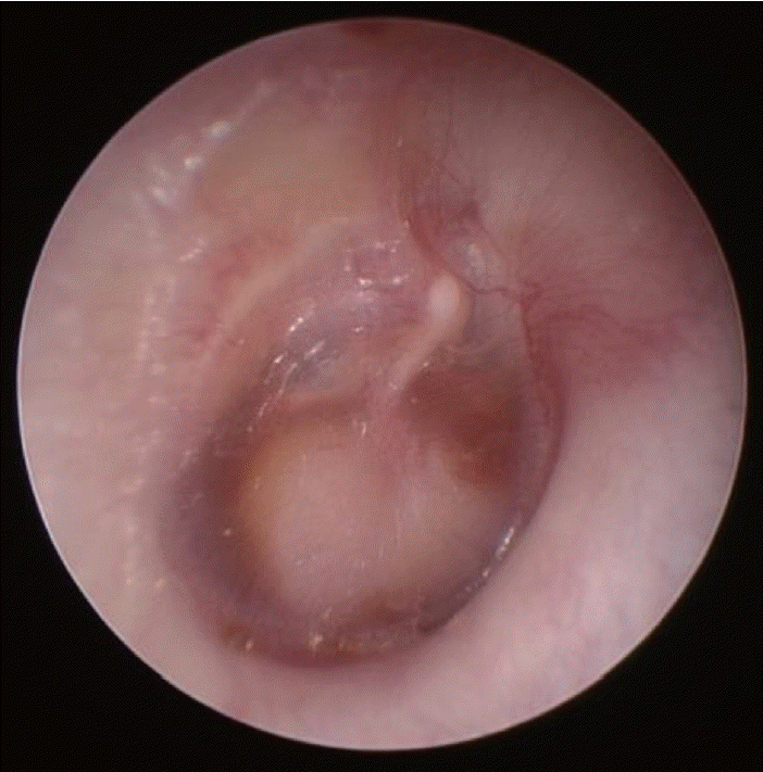
4세 10개월 나이의 여아가 우측 중이의 종물에 대한 진료를 위해 내원하였다. 아동은 1년 전부터 반복적으로 발생하는 우측 삼출성 중이염으로 거주지 인근 의원의 진료를 받던 중중이 종물이 의심되어 본원으로 전원되었다. 이내시경 검사에서 우측 고막의 중심부와 하방에 고막 전체 면적의 약 2/3를 차지하는 타원 형태의 백색 종물이 관찰되었다(Fig. 1). 순음청력검사에서 기도 청력(6분법 평균)은 우측 35 d B H L, 좌측 12 d B H L이었다. 환아가 차폐 검사를 수행하지 못해 골도청력검사는 시행하지 않았다. 이내시경 검사 소견에 근거하여 중이 진주종을 의심하고 측두골 전산화단층촬영을 시행하였다.
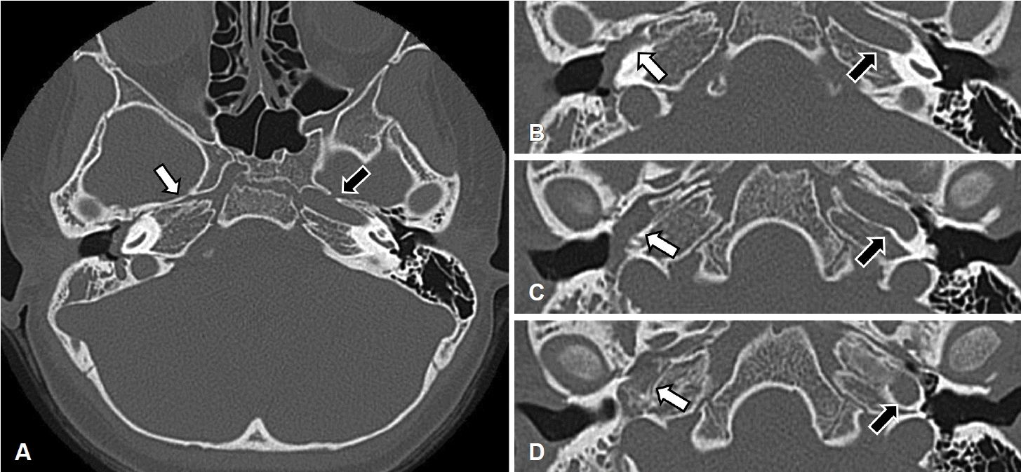
측두골 전산화단층촬영에서 우측 중고실, 하고실 전체와 이관의 입구부를 침범한 연조직 밀도의 병변이 관찰되었고 이소골 연쇄는 정상이었다. 동측 측두골 추체부에 내경동맥관이 결손되어 있었고, 중이 병변은 내경동맥 결손 부위 공간을 채우고 있었다(Fig. 2). 병변이 상고실을 제외한 중이강 전체를 침범하고 있으나 골결손과 이소골 연쇄의 손상이 없는 점에서 진주종이 아닌 다른 연부조직 종양 가능성이 제기되어 자기공명영상을 시행하였다.

The images of temporal bone CT. Axial images at the level of skull base are presented from superior (A) to inferior (D). The images show an absence of ICA at right side (white arrow) and an presence of normal ICA at left side (black arrow). Consecutive axial images from (B) to (D) show the soft tissue density lesion at right middle ear occupying the space where ICA should have been. ICA: internal carotid artery.
자기공명영상에서 우측 중이 병변의 침범부위는 전산화단층촬영의 소견과 동일하였고, 동측 내경동맥이 결손된 소견을 보였다. 중이 병변은 T1 강조영상에서 저신호강도, T2 강조영상에서 저신호강도, PROPELLA 시퀀스를 이용한 확산 강조영상에서 저신호강도를 보이고, Gd 조영증강 T1 강조영상에서 조영 증강되어 진주종보다는 연부조직 종양의 가능성을 시사하였다(Fig. 3).

MR images. T2-weighted image (A) shows a low signal intensity (arrow). Gadolinium enhanced T1-weighted image (B) shows an enhancing lesion (arrow).
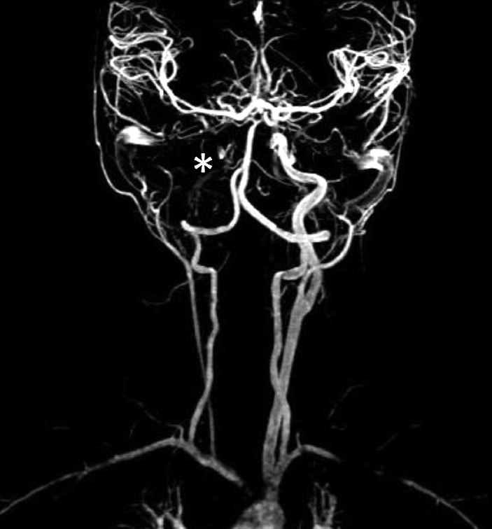
측두골 전산화단층촬영과 자기공명영상에서 우측 내경동맥이 결손된 소견을 보였기 때문에, 뇌혈류를 확인하기 위해 자기공명 혈관촬영을 추가로 시행하였다. 우측 내경동맥은 없었으나 좌측 내경동맥의 겉순환으로 인해 우뇌 반구 전 영역의 혈류 공급이 정상적으로 이루어지고 있었고, 동맥류와 같은 혈관 이상 소견은 없었다(Fig. 4).

The image of MR angiography. The right internal cervical artery (asterisk) is absent but ipsilateral anterior cerebral artery and middle cerebral artery are supplied by anterior communicating artery.
중이 종물의 제거를 위해 고실 개방술을 시행하였다. 후이개 절개를 가한 후 고실외이도 피판을 거상하였다. 중이 종물로 인한 압박으로 고막은 팽륜되어 있었으나 종물과 고막간 유착은 없었다. 종물은 중고실과 하고실의 대부분을 차지하고 있었는데, 종물이 크고 시야가 협소하여 종물의 전체를 확인할 수 없었다(Fig. 5). 종물은 세 이소골 모두와 접촉하고 있었으나 이소골의 미란은 없었다. 침골-등골 관절을 분리하여 침골을 제거하고 고막긴장건(tensor tympani tendon)을 자른 후 고실외이도 피판을 전방으로 완전히 밀어서 외이도 전벽에 닿도록 함으로써, 종물을 향한 시야와 종물을 제거할 작업 공간을 확보하였다. 종물은 전방 중고실(anterior mesotympanum)의 가장 깊은 부위에서 기시하여 중고실과 하고실로 자라 나온 양상이었다. 특이한 점은 내경동맥 무형성증으로 인해 전방 중고실이 전방으로 넓고 깊게 형성되어 있었으며, 깊게 형성된 전방 중고실의 가장 전방 부위에서부터 종물이 기시하고 있었다. 수술 현미경 시야를 통해 종물의 상당 부분을 제거하였으나, 종물이 기시한 전방 중고실의 가장 전방 부위는 수술 현미경 시야로는 접근되지 않아, 70° 내시경과 내시경 중이 수술 기구를 이용하여 전방 중고실에 위치한 종물을 완전제거하였다. 종물 제거 과정에서 고막과 고실륜의 결손이 발생하여, 측두근막을 이용하여 외면이식 고실성형술을 시행하고 수술을 마쳤다. 수술 후 합병증은 없었으며, 수술 이틀 후 퇴원하였다.

The surgical findings. (A) The microscopic view of right middle ear after elevating tympanomeatal flap shows large mass occupying mesotympanum and hypotympanum (asterisk). (B) The 30°endoscopic view after removing the posterior half of the mass. Anterior half of the mass (asterisk) is remained. Ossicular continuity is intact though long process of incus is bent. (C) The 70°endoscopic view showing anterior mesotympanum after further removing the mass. Tympanomeatal flap was pushed forward fully to be attached to anterior wall of external auditory canal after removing incus and cutting tensor tympani tendon. The remaining mass (asterisk) was attached tightly to anterior wall of enlarged anterior mesotympanum where ICA should had been. The last part of the mass is being removed using Fish dissector. The two dashed lines indicate the imaginary ICA. M: malleus, I: incus, S: stapes, FN: facial nerve, TM: tympanic membrane, TMF: tympanomeatal flap, ICA: internal carotid artery.
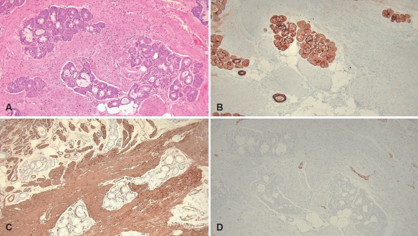
병리조직검사에서 종물은 정상 침샘 조직과 평활근, 지방세포, 말초신경 조직으로 구성되어 있었다. 면역조직화학염색에서 침샘 조직은 pan-CK, 평활근 조직은 smooth muscle actin, 말초신경 조직은 S100에 양성을 보여 과오종으로 확진하였다(Fig. 6). 수술 후 6개월 동안의 추적 관찰에서 고막은 치유되었고 내시경 진찰 소견상 재발 소견은 없으며, 추후 이소골 성형술을 시행할 예정이다.

The microscopic findings of the tumor. (A) The tumor was mainly composed of benign salivary gland, which were separated by smooth muscle bundles. The mature fat tissue and peripheral nerve bundles were interspersed (hematoxylin and eosin stain, ×100 magnification). (B) Immunohistochemical staining for pan-CK highlights the salivary gland (pan-CK, ×100 magnification). (C) The smooth muscle bundles were positive for SMA (SMA, ×100 magnification). (D) The peripheral nerve bundles showed positive immunoreactivity for S100 (S100, ×100 magnification). SMA: smooth muscle actin.
고 찰
과오종은 신체 모든 부위에서 발생할 수 있다. 과오종 자체에 의한 증상은 드물기 때문에 신체검사와 영상검사에서 우연히 발견되는 경우가 대부분이고, 드물게 증상이 있는 경우에는 종괴에 의한 압박증상이 나타날 수 있다[5]. 과오종의 유병률은 폐에서 발생한 경우를 제외하고는 밝혀져 있지 않다. 폐 과오종의 유병률은 약 0.25%로 모든 폐 종양의 약 8%를 차지한다[14]. 과오종의 조직병리적 특징은 연골과 혈관, 지방의 간엽조직과 상피, 분비선등의 상피성 조직이 혼재되고 특정 조직의 과도한 증식이 관찰되며 세포 이형성 또는 악성 변화는 극히 드물다[5]. 과오종은 완전절제가 가장 좋은 치료 방법이며, 완전절제한 경우 재발은 알려진 바가 없다[14].
중이 과오종은 발생 부위에 존재하는 정상 조직의 증식이라는 점에서 기형종(teratoma), 유피종(dermoid)과 감별하여야 한다. 기형종은 외배엽, 중배엽, 내배엽의 3배엽 모두에서 유래한 조직을 포함하고, 유피종은 외배엽과 중배엽에서 유래한 조직의 낭성 종괴라는 점에서 과오종과 구분된다. 또한 과오종은 분리종(choristoma)과도 감별하여야 하는데, 분리종은 정상적으로 해당부위에서 발견될 수 없는 조직이 증식하는 종양으로, 정상위치에서 정상적으로 존재하는 세포 중 일부가 비정상적인 성장과 배열을 보이는 과오종과 구분된다. 본 증례의 중이 종물은 평활근, 지방세포, 말초신경, 침샘 조직으로 구성되어 과오종으로 진단되었다. 비록 중이에 정상적으로 존재하지 않는 침샘 조직이 포함되었으나, 종물 전체가 침샘조직 단독으로 구성되지 않았고, 지방, 근육, 신경 조직 등 정상적으로 중이강에 존재하는 조직 성분이 종물의 대부분을 구성하였기 때문에 병리학적으로 과오종으로 진단되었다. 또한 중배엽과 외배엽에서 기원한 조직 성분만으로 구성되어 기형종도 배제할 수 있었다.
일측 내경동맥 무형성증은 매우 드문 선천성 혈관 기형으로 유병률은 약 0.01%이다. 일측 내경동맥 무형성증 환자는 대부분 무증상인데, 이는 윌리스 고리(Willis circle)의 겉순환으로 인해 결손된 내경동맥의 동측 대뇌반구의 모든 뇌실질에 정상적으로 혈액 공급이 이루어지기 때문이다[12]. 하지만 뇌동맥류 발생 가능성과 뇌혈류의 부전으로 인한 일과성 뇌허혈 발작의 발병률이 증가한다[13]. 내경동맥 무형성증 환자의 뇌동맥류 유병률은 25%~43%로 혈관기형이 없는 환자의 유병률 2%~4%보다 크게 높은 것으로 조사된 바 있다[12,13].
본 증례는 일측 내경동맥 무형성증을 가진 환자의 동측 중이에 발생한 과오종을 치험한 것으로 전 세계적으로 이와 같은 증례는 보고된 바가 없다. 본 증례 환자는 내경동맥 무형성증으로 인한 뇌혈관 관련 문제는 없었으나, 내경동맥의 결손으로 인해 전방 전고실이 매우 깊게 형성되어 있었고, 이 공간의 가장 깊숙한 부위에서 과오종이 발생하여 시야 확보와 종물 제거에 어려움이 있었다.
정상 고막을 가진 소아에서 중이 종양이 의심될 때 선천성 진주종, 콜레스테롤 육아종, 안면신경종, 횡문근육종, 육종, 지방종, 기형종 등을 감별해야 한다[15]. 소아에서 중이 내에 백색에 가까운 종물이 보일 경우 선천성 진주종의 가능성이 가장 높으나 본 증례에서 보듯이 과오종의 가능성도 염두에 둘 필요가 있다는 점과, 내경동맥의 결손은 뇌동맥류를 포함한 뇌혈관 문제 외에도 중이 공간의 변형을 초래할 수 있다는 두가지 임상적 의의가 있어 본 증례를 보고하는 바이다.
Acknowledgements
None.
Notes
Author Contribution
Conceptualization: Sung Wook Jeong. Data curation: Sangjun Kim. Methodology: Sung Wook Jeong. Visualization: Min Gyoung Pak. Writing—original draft: Sangjun Kim. Writing—review & editing: Sung Wook Jeong.

