비전정 협착을 수술적으로 치료한 새로운 기법 1예
A Case of Surgical Correction of Nasal Vestibular Stenosis
Article information
Trans Abstract
Nasal vestibular stenosis is an uncommon cause of nasal obstruction. There are various causes of the narrowing of vestibule, including previous trauma, surgery, infection, and iatrogenic injury. The disease is characterized by circumferential scar retraction in inlet of nasal cavities which are caused by secondary proliferation of granulation and fibrous tissue. A number of studies were reported on the surgical correction of vestibular stenosis, but no single technique had been widely accepted due to a wide spectrum treatment methods owing to the various causes of disease and deformities of the related anatomy. Here we suggest a new surgical technique for correcting a case of vestibular stenosis caused by trauma.
서 론
비전정 협착(vestibular stenosis)은 코막힘을 유발하는 드문 질환으로 다양한 원인에 의해 발생할 수 있지만, 크게 선천적 협착과 후천적 협착으로 분류할 수 있다. 이중, 후천적 협착은 주로 외상, 감염, 화상, 과거 수술로 인한 반흔 등에 의해 유발되는 것으로 알려져 있다[1]. 이러한 협착이 발생하는 병리학적 특징은 비전정 피부 밑 피하조직의 손상, 특히 윤상의 상처(circumferential scar), 발생 이후 2차적인 섬유화 조직의 증식이다[2,3].
과거의 다양한 연구자들이 이를 치료하기 위한 방법들을 고안해 내고 제시하고 있으나 표준적인 치료가 확립되어 있지는 않다. 이는 환자마다 질병의 발생원인 및 동반된 해부학적 구조의 변형이 달라 치료의 범위가 다양하기 때문일 것으로 생각된다. 일부에서는 수술 이후 스텐트 등의 보조 요법이 도움이 된다고 보고하고 있으나, 그럼에도 불구하고 불충분한 교정과 잦은 재발로 인해 치료가 어려운 것으로 알려져 있다[4-6].
본 연구에서는 반흔 조직의 절제 및 비강 내 국소 피판을 이용하여 성공적으로 비전정 협착을 치료한 증례를 소개하고자 한다.
증 례
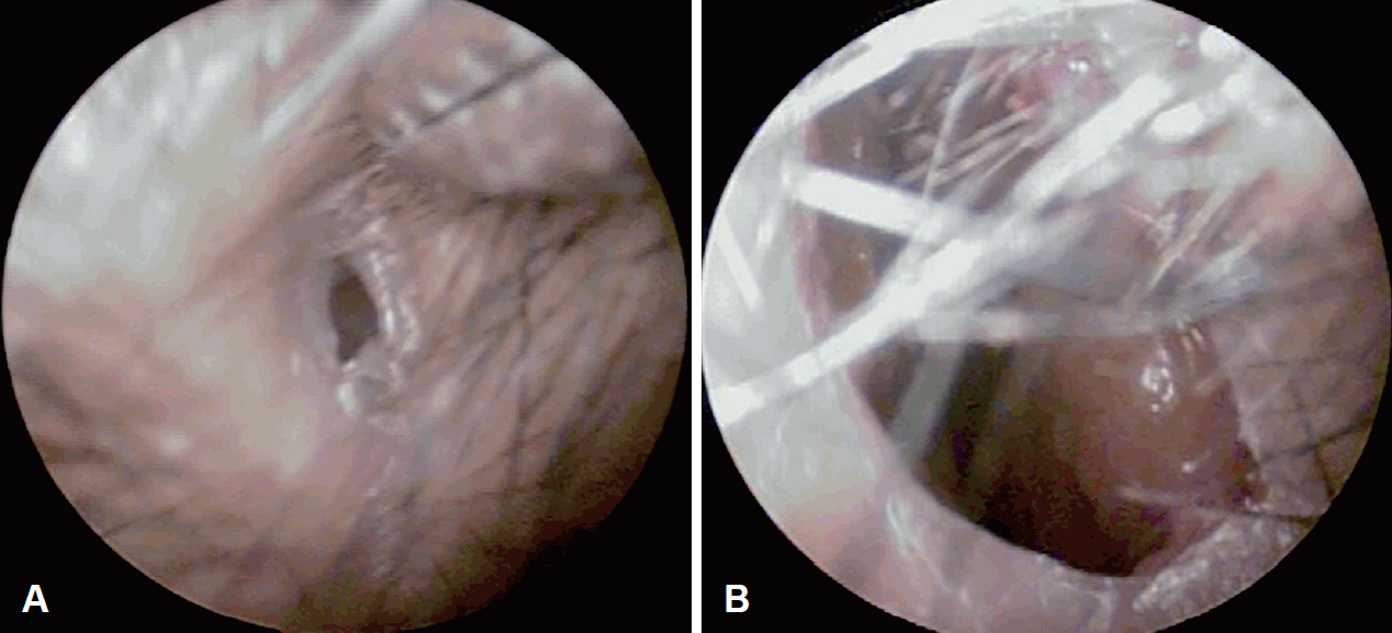
52세 여자 환자가 좌측 코막힘을 호소하며 내원하였다. 과거력상 어린 시절 외상력이 있었으며, 30대에 미용적 목적의 코성형술을 시행받았다. 진찰 소견에서 환자는 좌측 비익의 경미한 함몰과 함께 동측의 비전정부의 협착 소견을 보였다(Fig. 1). 외비강의 90% 이상을 폐쇄하고 있는 협착부로 인해 비강 내부의 소견을 확인할 수 없었으므로, 추가적인 평가를 위하여 시행한 전산화단층촬영에서 이와 동일한 소견과 함께 주변의 외비를 구성하는 연골 등의 지지 구조나 비강, 부비동에는 이상 소견이 없음을 확인할 수 있었다(Fig. 2).

Preoperative endoscopic findings of both nasal vestibules. Right nasal cavity (A), left nasal cavity (B).
수술적인 교정을 위해 상담을 시행하였을 때, 환자는 추가적인 미용적 수술은 원하지 않고 기능적 개선만을 원하여 코막힘을 개선하기 위해 수술을 시행하였다. 수술은 전신마취하에 진행되었으며, 수술의 과정을 순서대로 요약하자면, 협착부의 11시와 4시에 절개를 가하고 외부의 피부 피판을 거상한 이후(Fig. 3A), 비강 측(asterisk of Fig. 3B)의 협착 부위는 충분히 절제하여 주었다. 이후 절제가 시행된 연조직 노출 부위에 비강 외측의 피부피판을 덮어주고 silastic sheet를 덮어주었다(Fig. 3B). Silastic sheet는 비전정을 모두 덮어줄 수 있게 디자인하여 거치 및 봉합 고정한 이후 수술 후 3주에 제거하였다.

Schematic diagram of the technique used in cases. Superior-medial and inferior-lateral incision was made first for obtaining sufficient surgical field. Perpendicular incisions were made by using crescent knife at the edge of stenotic segment. After then, two skin flaps of nasal vestibule were elevated to expose the stenotic segment (A). Cross-sectional view; outer skin flaps were elevated, after then resection of fibrotic tissues in inner stenotic portion (nasal cavity side; asterisk, *) and repositioning of the outer flaps (vestibular skin) were performed for preventing restenosis (B).
환자는 수술 후 경과에서 2년 이상 재협착 없이 성공적으로 피판이 상피화되었으며, 주관적으로 코막힘 증상도 해소되었다(Fig. 4).
고 찰
비전정 협착은 드문 질환이며, 일부의 치험과 증례만이 국내외의 학술지에 소개가 되고 있다. 이는 질병 자체가 다양한 원인에 의해 발생하는 질환이어서, 각 증례가 가진 특징과 동반된 변형에 따라 여러 복합적인 치료 방침을 적용하기 때문으로 생각된다. 본 증례 보고에서 저자들은 다른 해부학적 변이가 심하지 않은 경우에 활용할 수 있는 술식을 소개하였다.
비전정 협착은 해부학적 위치상 외비밸브 기능의 저하를 필연적으로 동반하기 때문에 대부분의 환자는 코막힘을 주증상으로 하며, 협착부의 정도에 따라 외비의 변형을 동반할 수 있다. 진단과 치료 방침 결정을 위해서는 병변의 원인이 되는 과거력 및 수술력 등을 포함한 임상 정보를 세밀하게 평가하여야 함은 물론이고, 전산화단층촬영을 통해 동반된 해부학적 변형의 여부를 평가할 수 있고, 또한 협착부의 위치와 정도를 평가할 수 있다. 본 증례에서는 비중격 혹은 비강 내의 다른 복합적인 변형없이 비전정 부위에 국한된 협착 부위를 확인할 수 있었다.
비전정 협착의 수술적 치료는 아직 표준 치료가 정립되지 않았고 치료 방침 설정과 수술의 난이도가 높은 것으로 알려져 있다. 협착의 정도와 함께 주변 해부학적 구성요소들의 변형 정도에 따라 다양한 방법의 수술 방법들을 적용할 수 있는데, Z-plasty나 W-plasty에서부터 국소피판, 복합조직편의 이식 등이 이에 해당한다[4,5,7-9]. 수술의 범위가 커질수록 수술 후의 재협착이나 치료 실패의 가능성도 높아지는 경향이 있으므로, 일부 저자들은 수술 후 치료의 중요성을 강조하고 있으며 비강 내 장치들을 제안하기도 한다[3,6].
질병의 발생이 피부와 그 부속 기관에 발생한 상처의 치유 과정에서 발생하는 과도한 섬유화가 원인이 되므로, 수술 시에도 이를 고려하여야 한다[1,2]. 비전정 협착 수술의 원칙은 충분한 수술 전 계획과 수술 중의 세심한 박리 및 절제이다. 특히 수술 중 박리 및 절제는 수술 후 재협착을 유발하는 섬유화를 최소화하기 위한 노력이 필요하다. 본 증례에서 소개하는 술식은 지나친 절제를 최소화할 수 있으면서도, 비강 내 접근을 통해 비전정의 피부-점막 이행부의 단순 원형 절개 및 피부 견인을 통한 피판 거상을 통해 시행할 수 있어, 기존에 소개된 디자인이 다소 어려울 수 있는 Z-plasty나 W-plasty에 비해 장점을 지닌다 하겠다. 하지만 다양한 변형이 동반되거나 연조직 외의 연골부 변형 등의 복합적 변형의 교정에서의 효과는 제한적일 것으로 생각된다.
본 저자들은 이 증례 보고를 통하여 비전정에 발생한 해부학적 협착이 복합적이지 않은 경우에는 반흔 조직의 충분한 절제 및 비내 피부 피판을 이용한 교정이 효과적인 선택이 될 수 있다고 제안하고자 한다. 본 술식의 경우 기존의 술식보다 간편하면서도 재협착의 방지에도 효과적인 새로운 술식이며, 추후 추가적인 증례가 보완된다면 비전정 협착의 치료에 더욱 유용한 수술 방법이 될 것으로 생각한다.
Acknowledgements
None.
Notes
Author Contribution
Conceptualization: all authors. Formal analysis: Yung Jin Jeon, Sang-Wook Kim. Funding acquisition: Yung Jin Jeon, Sang-Wook Kim. Investigation: Sang-Wook Park, Yung Jin Jeon, Sang-Wook Kim. Methodology: Hyun-Jin Cho. Resources: Sang-Wook Park, Hyun-Jin Cho. Supervision: Yung Jin Jeon, Sang-Wook Kim. Validation: Yung Jin Jeon, Sang-Wook Kim. Visualization: Hyun-Jin Cho. Writing—original draft: Sang-Wook Park. Writing—review & editing: Yung Jin Jeon, Sang-Wook Kim, Hyun-Jin Cho.


