 |
 |
AbstractCervical spondylosis is a common degenerative disease of the cervical spine affecting the cervical vertebral bodies and intervertebral discs. During parotidectomy, the patient is placed in a supine position with the neck extended and head rotated to the contralateral side. This position could exacerbate pre-existing cervical spondylosis and cause cervical myelopathy. We present a case of postoperative quadriplegia secondary to cervical myelopathy after parotidectomy. A 68-year-old man without symptoms of cervical spondylosis underwent partial parotidectomy for a right parotid mass and subsequently developed quadriplegia 8 hours postoperatively. Magnetic resonance imaging revealed severe cervical myelopathy. Emergency laminoplasty was performed, and steroid therapy was initiated. He showed near-complete recovery six months later.
м„ң лЎмқҙн•ҳм„ м Ҳм ңмҲ (parotidectomy)мқҖ л‘җкІҪл¶Җмҷёкіј мҳҒм—ӯм—җм„ң нқ”нһҲ мӢңн–үлҗҳлҠ” мҲҳмҲ мқҙлӢӨ. мқҙн•ҳм„ м Ҳм ңмҲ нӣ„ л°ңмғқн• мҲҳ мһҲлҠ” лҢҖн‘ңм Ғмқё н•©лі‘мҰқмңјлЎңлҠ” м–јкөҙ мӢ кІҪ л§Ҳ비, Freyм”Ё мҰқнӣ„кө°, н”јнҢҗ кҙҙмӮ¬, мһ¬л°ң л“ұмқҙ мһҲлӢӨ. мқҙн•ҳм„ м Ҳм ңмҲ мқҖ лҢҖк°ң м•ҷмҷҖмң„ мһҗм„ём—җм„ң мҲ мёЎмқҳ л°ҳлҢҖнҺёмңјлЎң лЁёлҰ¬лҘј нҡҢм „мӢңнӮӨкі лӘ©мқ„ мӢ м „мӢңнӮЁ мһҗм„ёлЎң мӢңн–үлҗңлӢӨ. мқҙ мһҗм„ёлҠ” мһ мһ¬м ҒмңјлЎң кё°мЎҙмқҳ кІҪ추 мІҷ추мҰқ(cervical spondylosis)мқ„ м•…нҷ”мӢңнӮ¬ мҲҳ мһҲкі кІҪ추 мІҷмҲҳлі‘мҰқ(cervical myelopathy)мқ„ мҙҲлһҳн• мҲҳ мһҲлӢӨ[1-3]. мқҙн•ҳм„ м Ҳм ңмҲ л“ұ л‘җкІҪл¶Җмҷёкіј мҲҳмҲ нӣ„ л°ңмғқн•ң мӮ¬м§Җл§Ҳ비лҠ” м•„м§Ғ көӯлӮҙм—җм„ң ліҙкі лҗң л°”к°Җ м—Ҷм—ҲлӢӨ. м Җмһҗл“ӨмқҖ кІҪ추 мІҷ추мҰқмқ„ 진лӢЁл°ӣм§Җ м•ҠмқҖ нҷҳмһҗм—җм„ң мқҙн•ҳм„ м Ҳм ңмҲ нӣ„ кІҪ추 мІҷмҲҳлі‘мҰқмңјлЎң мқён•ң мӮ¬м§Җл§Ҳ비 мҰқлЎҖлҘј кІҪн—ҳн•ҳмҳҖкё°м—җ л¬ён—Ң кі м°°кіј н•Ёк»ҳ ліҙкі н•ҳкі мһҗ н•ңлӢӨ.
ліё м—°кө¬лҠ” к°•лҰүм•„мӮ°лі‘мӣҗ кё°кҙҖмғқлӘ…мңӨлҰ¬мң„мӣҗнҡҢ(Institutional Review Board)мқҳ мҠ№мқёмқ„ л°ӣм•ҳлӢӨ.
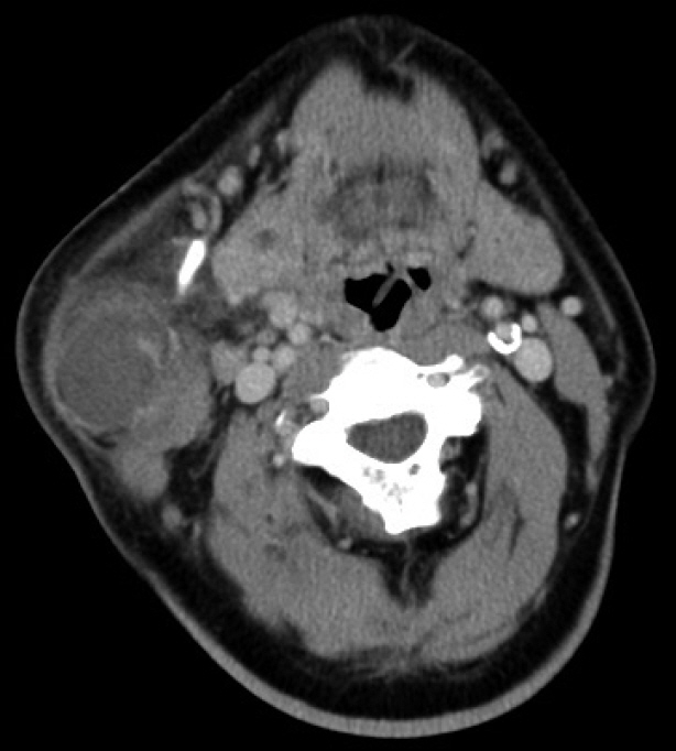
мҰқ лЎҖ68м„ё лӮЁмһҗк°Җ 5л…„ м „л¶Җн„° мһҗк°Ғн•ҳкі мһҲлҚҳ мҡ°мёЎ мқҙн•ҳм„ л¶Җмң„ мў…кҙҙк°Җ лӮҙмӣҗ 3мқј м „л¶Җн„° кёүкІ©н•ҳкІҢ нҒ¬кё°к°Җ мҰқлҢҖлҗҳм–ҙ мҷёлһҳм—җ лӮҙмӣҗн•ҳмҳҖлӢӨ. мӢ мІҙкІҖмӮ¬м—җм„ң мҳӨлҘёмӘҪ мқҙн•ҳм„ л¶Җмң„м—җ м••нҶө, м—ҙк°җмқҙ лҸҷл°ҳлҗң 10 cm нҒ¬кё°мқҳ мў…кҙҙк°Җ кҙҖм°°лҗҳм—ҲлӢӨ. кІҪл¶Җ м»ҙн“Ён„°лӢЁмёөмҙ¬мҳҒмқ„ мӢңн–үн•ҳмҳҖмңјл©°, мҡ°мёЎ мқҙн•ҳм„ мІңм—Ҫл¶Җмң„ мЈјліҖм—җ мЎ°мҳҒмҰқк°•мқ„ ліҙмқҙлҠ” 3.5 cmмқҳ мў…л¬јмқҙ кҙҖм°°лҗҳм—Ҳмңјл©°, мЈјліҖ н”јн•ҳм§Җл°©мёөмңјлЎң м—јмҰқмқҙ нҢҢкёүлҗҳм–ҙ мһҲлҠ” мҶҢкІ¬мқҙм—ҲлӢӨ(Fig. 1). мҲ м „ мӮ¬м§Җмқҳ к·јл ҘмқҖ м •мғҒмқҙм—Ҳкі мӢ кІҪн•ҷм Ғ мҰқмғҒмқҖ нҳёмҶҢн•ҳм§Җ м•Ҡм•ҳлӢӨ. мҡ°мёЎ мІңм—Ҫм Ҳм ңмҲ (right partial superficial parotidectomy)мқ„ мӢңн–үн•ҳмҳҖкі , лі‘лҰ¬мЎ°м§Ғн•ҷм ҒмңјлЎң мҷҖлҘҙнӢҙ мў…м–‘(warthin tumor)мңјлЎң 진лӢЁлҗҳм—ҲлӢӨ. мҙқ мҲҳмҲ мӢңк°„мқҖ 4мӢңк°„к°Җлҹү мҶҢмҡ”лҗҳм—ҲлӢӨ. мҲҳмҲ м§Ғнӣ„ кІҪлҜён•ң лӘ© нҶөмҰқмқ„ нҳёмҶҢн•ҳмҳҖкі , нҷҳмһҗлҠ” мҲҳмҲ нӣ„ 8мӢңк°„ л§Ңм—җ мӮ¬м§Җл§Ҳ비лҘј мІҳмқҢмңјлЎң ліҙкі н•ҳмҳҖлӢӨ. к·јл Ҙ нҸүк°Җ кІ°кіј м–‘мёЎ мғҒм§Җ 1/5, м–‘мёЎ н•ҳм§Җ 1/5мқҳ нҳ„м Җн•ң к·јл Ҙ м Җн•ҳк°Җ кҙҖм°°лҗҳм—ҲлӢӨ. к·ёлҹ¬лӮҳ мқҳмӢқ ліҖнҷ”, к°җк°Ғ мһҘм• л“ұмқҳ лӢӨлҘё мӢ кІҪн•ҷм Ғ мҰқмғҒмқҖ кҙҖм°°лҗҳм§Җ м•Ҡм•ҳлӢӨ.
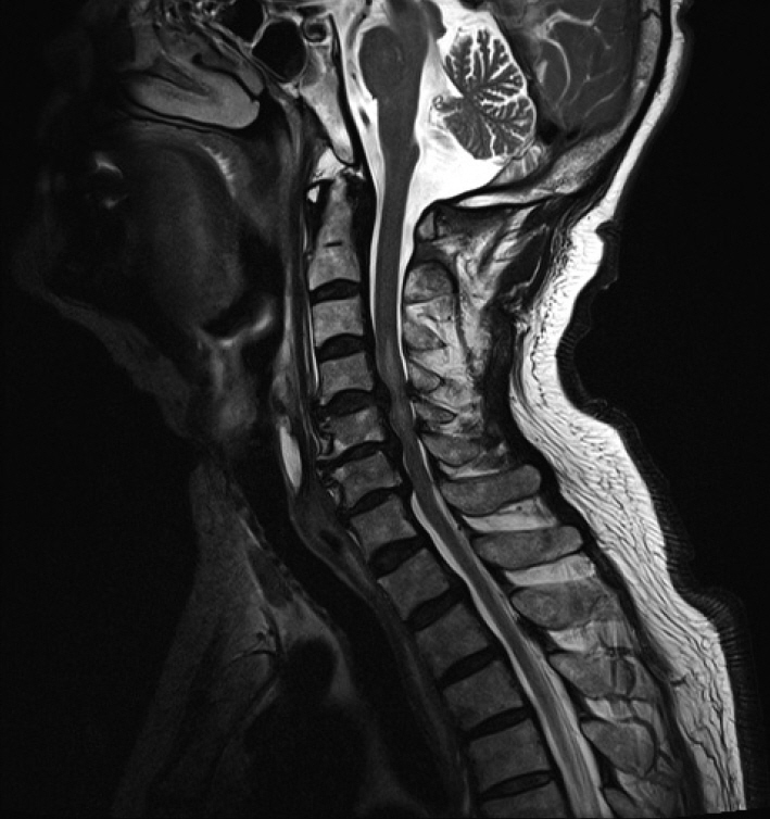
мҰүк°Ғм ҒмңјлЎң мӢ кІҪмҷёкіј нҳ‘진мқ„ мқҳлў°н•ҳм—¬ кІҪ추 мһҗкё°кіөлӘ…мҳҒмғҒмқ„ мӢңн–үн•ҳмҳҖмңјл©° T2 к°•мЎ° мҳҒмғҒм—җм„ң м ң4-5 кІҪ추간л¶Җн„° м ң6-7 кІҪ추간м—җ мқҙлҘҙлҠ” мӢ¬н•ң мІҷ추 нҳ‘м°©(spinal stenosis)мқ„ лҸҷл°ҳн•ң м••л°•м„ұ мІҷмҲҳлі‘мҰқ(compressive myelopathy)мқҙ кҙҖм°°лҗҳм—ҲлӢӨ(Fig. 2). мҰқмғҒ нҳёмҶҢ 10мӢңк°„ нӣ„ м ң4-5-6 кІҪ추мқҳ нҺёмёЎ нӣ„к¶Ғмқ„ м—ҙм–ҙ мӢ кІҪкҙҖмқ„ нҷ•мһҘмӢңнӮӨлҠ” мўҢмёЎ к°ңл°©л¬ё 추к¶Ғ м„ұнҳ•мҲ (left open door laminoplasty)мқ„ мӢңн–үн•ҳмҳҖкі , л©”нӢён”„л Ҳл“ңлӢҲмҶ”лЎ (methylprednisolone) 125 mg м •л§Ҙ лӮҙ мқјмӢңмЈјмӮ¬ нӣ„ л§Ө 6мӢңк°„л§ҲлӢӨ 60 mg м •л§Ҙ лӮҙ мЈјмӮ¬лЎң 48мӢңк°„ лҸҷм•Ҳ мҠӨн…ҢлЎңмқҙл“ң м№ҳлЈҢ(steroid therapy)лҘј мӢңн–үн•ҳмҳҖлӢӨ. к·јл Ҙ нҡҢліөмқҖ м–‘нҳён•ҳм—¬ мҲҳмҲ 3мқј нӣ„ лӘЁл“ мӮ¬м§Җмқҳ к·јл ҘмқҖ 4/5к№Ңм§Җ нҡҢліөлҗҳм—ҲлӢӨ. 추к¶Ғ м„ұнҳ•мҲ мӢңн–ү 100мқј нӣ„ нҷҳмһҗлҠ” нҮҙмӣҗн•ҳмҳҖкі , м•Ҫ 2л…„ 1к°ңмӣ”мқҙ м§ҖлӮң нҳ„мһ¬ нҷҳмһҗлҠ” кІҪлҜён•ң кі мң мҲҳмҡ©м„ұ к°җк°Ғ м Җн•ҳл§Ңмқ„ ліҙмқҙл©° мҲҳмҲ м „ к·јл Ҙм—җ к·јм ‘н•ҳкІҢ нҡҢліөлҗҳм–ҙ м§Җм—ӯмӮ¬нҡҢ ліҙн–үмқҙ к°ҖлҠҘн•ң мғҒнғңмқҙлӢӨ.
кі м°°мӨ‘мҰқмқҳ кІҪ추 мІҷмҲҳлі‘мҰқмқҖ мқҙн•ҳм„ м Ҳм ңмҲ мқҙлӮҳ кё°нғҖ л‘җкІҪл¶Җмҷёкіј мҲҳмҲ м—җм„ң л§Өмҡ° л“ңл¬ё н•©лі‘мҰқмқҙлӢӨ. 비мІҷмҲҳмҲҳмҲ мқё л‘җкІҪл¶Җ мҲҳмҲ нӣ„ мІҷмҲҳ мҶҗмғҒмқҖ мқҙм „м—җ к°‘мғҒм„ мҲҳмҲ м—җм„ң ліҙкі лҗң л°”к°Җ мһҲм—ҲлӢӨ[4]. кІҪ추 мІҷмҲҳлі‘мҰқмқҳ лі‘нғңмғқлҰ¬лҠ” м•„м§Ғ л¶Ҳ분лӘ…н•ҳлӮҳ мІҷмҲҳн—ҲнҳҲ, кІҪл§үмҷё нҳҲмў…, м§Ғм ‘м Ғмқё м••л°• лҳҗлҠ” кІ¬мқё л“ұмқ„ мӣҗмқёмңјлЎң м¶”м •н•ҳкі мһҲлӢӨ.
кІҪ추 мІҷмҲҳлі‘мҰқмқҖ кІҪ추мқҳ л…ёнҷ”м—җ мқҳн•ҙ л°ңмғқн•ҳлҠ” 진н–үм„ұ лі‘ліҖ мӨ‘ к°ҖмһҘ нқ”н•ҳлӢӨ. мқҙ м§ҲнҷҳмқҖ кіЁмҰқмӢқм„ұкіЁк·№(osteophytic spur)мқҳ л°ңмғқмқҙлӮҳ, мІҷ추мӮ¬мқҙмӣҗл°ҳ(intervertebral disc)мқҳ 붕кҙҙ л°Ҹ лҸҢм¶ң, к·ёлҰ¬кі нҷ©мғү мқёлҢҖ(ligamentum flavum)мқҳ 비лҢҖлЎң мІҷ추кҙҖмқҙ мўҒм•„м§ҖлҠ” кІғмқҙ нҠ№м§•мқҙлӢӨ.
мҰқмғҒмқҙ м—ҶлҠ” 60м„ё мқҙмғҒмқҳ мқёкө¬мқҳ м•Ҫ 85%м—җм„ң лӢӨм–‘н•ң м •лҸ„мқҳ л””мҠӨнҒ¬ ліҖм„ұмқҙ кҙҖм°°лҗңлӢӨ[5]. л‘җкІҪл¶Җмҷёкіј мҲҳмҲ м—җм„ң мһҗмЈј мӢңн–үлҗҳлҠ” лӘ©мқҳ мӢ м „мқҖ кё°мЎҙмқҳ л¬ҙмҰқмғҒ кІҪ추 мІҷ추мҰқмқ„ мӢ мІҙм Ғ кё°лҠҘ м Җн•ҳк°Җ лҸҷл°ҳлҗң мІҷ추мҰқмңјлЎң 진н–үмӢңнӮ¬ мҲҳ мһҲлӢӨ. лӘ© мӢ м „мқҖ нҷ©мғү мқёлҢҖмқҳ көҙкіЎкіј мІҷмҲҳ нӣ„л°© 종축 л°©н–Ҙ нҳҲкҙҖмқҳ көҙкіЎмқ„ м•јкё°н•ҳм—¬ мІҷмҲҳлҘј л“ұ мӘҪм—җм„ң м••л°•н• мҲҳ мһҲлӢӨ[6]. Brieg л“ұ[7]кіј Taylor [8]лҠ” кіЁмҰқмӢқм„ұ кіЁк·№мқҙ мһҲмқ„ л•Ң кіјмӢ м „м—җ мқҳн•ҙ мІҷмҲҳмқҳ мӢ¬н•ң м••л°•мқҙ л°ңмғқн• мҲҳ мһҲлӢӨкі ліҙкі н•ҳмҳҖлӢӨ. л°ҳл©ҙм—җ мёЎл©ҙ көҪнһҳ л°Ҹ 축 нҡҢм „мқҖ мӢ м „м—җ 비н•ҳм—¬ мІҷмҲҳм—җ м ҒмқҖ мҳҒн–Ҙмқ„ лҜём№ңлӢӨ[6]. лҳҗн•ң мІҷ추кҙҖ нҳ‘м°©мқҖ көҙкіЎмӢң ліҙлӢӨ мӢ м „мӢңм—җ л‘җ л°° мҰқк°Җн•ҳлҠ” кІғмқҙ кҙҖм°°лҗҳм—ҲлӢӨ[9]. мқҙлҹ¬н•ң кё°м „мңјлЎң, лӘ©мқ„ мӢ м „мӢңнӮЁ мһҗм„ёлЎң мһҘмӢңк°„ мӢңн–үлҗҳлҠ” мҲҳмҲ мқҖ кІҪ추 мІҷмҲҳлі‘мҰқ мҰқмғҒмқ„ мң л°ңн• мҲҳ мһҲлӢӨ.
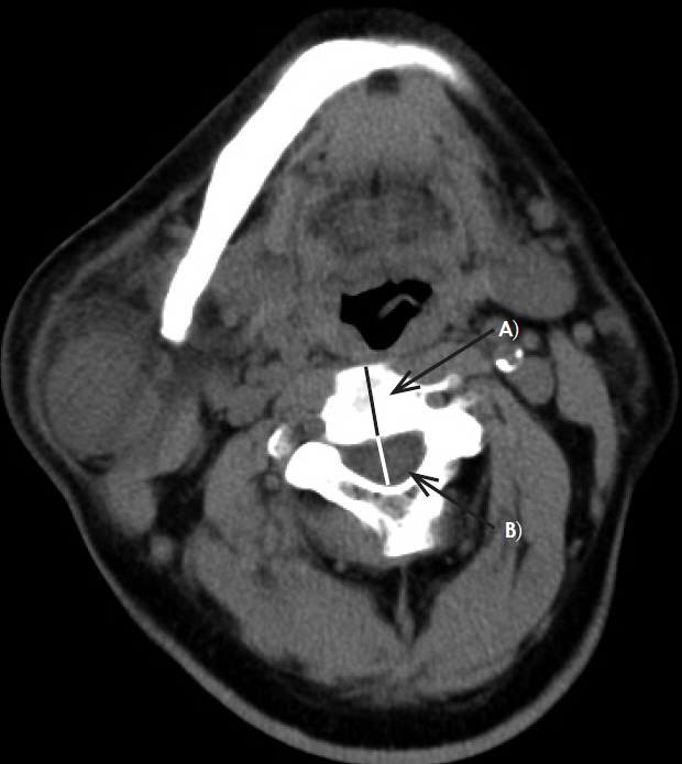
мҲҳмҲ нӣ„ мІҷмҲҳ мҶҗмғҒ к°ҖлҠҘм„ұмқ„ л‘җкІҪл¶Җмҷёкіј мҲҳмҲ м „ нҶөмғҒм ҒмңјлЎң мӢңн–үн•ҳлҠ” м»ҙн“Ён„°лӢЁмёөмҙ¬мҳҒмқҙлӮҳ мһҗкё°кіөлӘ…мҳҒмғҒмқ„ нҶөн•ҳм—¬ мҳҲмёЎн• мҲҳ мһҲлӢӨ[10]. мІҷ추кҙҖ м „нӣ„кІҪ(anteroposterior diameter of spinal canal)мқҙ 비көҗм Ғ л„“мқҖ м ң2-3 кІҪ추간 мІҷмҲҳмӮ¬мқҙмӣҗл°ҳ лҶ’мқҙм—җм„ң мІҷ추кҙҖ м „нӣ„кІҪкіј мІҷ추мӮ¬мқҙмӣҗл°ҳмқҳ м „нӣ„кІҪ 비(Torg-Pavlov ratio)к°Җ 0.59 лҜёл§Ңмқё нҷҳмһҗ, мІҷ추кҙҖ м „нӣ„кІҪмқҙ 8 mm лҜёл§Ңмқё мһҗ, мӢ мІҙкІҖмӮ¬мғҒ кІҪл¶ҖлҘј нҺёмёЎмңјлЎң көҪнһҢ нӣ„ лЁёлҰ¬ мң„мӘҪм—җм„ң м••л Ҙмқ„ к°Җн•ҳлҠ” мҠӨнҺ„л§Ғ кІҖмӮ¬ м–‘м„ұ нҷҳмһҗлҠ” мҲ м „ кІҪ추м—җ лҢҖн•ң нҸүк°Җк°Җ н•„мҡ”н•ҳлӢӨ(Fig. 3) [11-13].
ліё мҰқлЎҖмқҳ нҷҳмһҗмҷҖ к°ҷмқҖ кёүм„ұ мІҷмҲҳ мҶҗмғҒ(acute spinal cord injury)мңјлЎң мқён•ң л¶Ҳмҷ„м „ мӮ¬м§Җл§Ҳ비(incomplete quadriplegia)лЎң мһ¬нҷң м№ҳлЈҢлҘј л°ӣмқҖ нҷҳмһҗ мӨ‘ 21%мқҳ нҷҳмһҗк°Җ 6к°ңмӣ” нӣ„ ліҙн–ү лҠҘл Ҙмқ„ нҡҚл“қн•ҳм§Җ лӘ»н–ҲлӢӨлҠ” ліҙкі к°Җ мһҲлӢӨ[14]. лҳҗн•ң мІҷмҲҳ мҶҗмғҒ нҷҳмһҗлҠ” мһҘкё°м ҒмңјлЎң нҸҗл ҙ л“ұмқҳ нҳёнқЎкё°кі„ м§Ҳнҷҳкіј мһҗмңЁмӢ кІҪ л°ҳмӮ¬л¶Җм „(autonomic dysreflexia) л“ұмқҳ мӢ¬нҳҲкҙҖкі„ м§Ҳнҷҳмқ„ 비лЎҜн•ҳм—¬ л°©кҙ‘ кё°лҠҘл¶Җм „(bladder dysfunction), к·јмңЎ кІҪм§Ғ(spasticity), нҶөмҰқ мҰқнӣ„кө°(pain syndrome)мқҙлӮҳ м••л°•к¶Өм–‘(pressure ulcer)кіј к°ҷмқҖ лӢӨм–‘н•ң л§Ңм„ұм Ғ н•©лі‘мҰқмқҙ лі‘л°ңн• мҲҳ мһҲлӢӨ[15].
к·ёлҹ¬лҜҖлЎң мҲҳмҲ нӣ„ л°ңмғқн•ҳлҠ” мӮ¬м§Җл§Ҳ비лҠ” мЎ°кё° л°ңкІ¬кіј мҰүк°Ғм Ғмқё мӨ‘мһ¬к°Җ л§Өмҡ° мӨ‘мҡ”н•ҳлӢӨ. кёүм„ұ мІҷмҲҳ мҶҗмғҒм—җм„ң мҠӨн…ҢлЎңмқҙл“ң м№ҳлЈҢмҷҖ мҲҳмҲ м Ғ мӨ‘мһ¬ мӢңкё°м—җ лҢҖн•ҳм—¬ лӢӨмҶҢ л…јлһҖмқҙ мһҲм§Җл§Ң мІҷмҲҳ мҶҗмғҒ л°ңмғқ нӣ„ к°ҖлҠҘн•ҳл©ҙ 8мӢңк°„ мқҙлӮҙм—җ мҠӨн…ҢлЎңмқҙл“ң мҡ”лІ•мқ„ мӢңмһ‘н•ҳкі 24мӢңк°„ мқҙлӮҙм—җ мҲҳмҲ м Ғ мӨ‘мһ¬лҘј мӢңн–үн•ҳлҠ” кІғмқҙ мўӢлӢӨ[16,17]. ліё мҰқлЎҖмқҳ нҷҳмһҗлҠ” мҰүк°Ғм Ғмқё м№ҳлЈҢлЎң мҲҳмҲ м „ к·јл Ҙм—җ к·јм ‘н•ҳкІҢ нҡҢліөлҗ мҲҳ мһҲм—ҲлӢӨ.
л‘җкІҪл¶Җ мҲҳмҲ м—җ мһҲм–ҙм„ң кІҪ추 мІҷмҲҳлі‘мҰқмқҖ л“ңл¬јм§Җл§Ң л§Өмҡ° м№ҳлӘ…м Ғмқё н•©лі‘мҰқмқҙлӢӨ. мһҘмӢңк°„ лӘ©мқҳ мӢ м „мқҙ н•„мҡ”н•ң л‘җкІҪл¶Җ мҲҳмҲ мқ„ мӢңн–үн•ҳлҠ” кі л № нҷҳмһҗлҠ” мҲҳмҲ м „ кІҪ추 мІҷ추мҰқ 징нӣ„м—җ лҢҖн•ң мғҒм„ён•ң нҸүк°Җк°Җ мқҙлЈЁм–ҙм ём•ј н•ңлӢӨ. лҳҗн•ң мҲҳмҲ мӨ‘ лӘ©мқҳ мһҗм„ё ліҖнҷ”мҷҖ кіјмӢ м „мқҖ мөңмҶҢнҷ”лҗҳм–ҙм•ј н•ҳл©° н•©лі‘мҰқмқҙ л°ңмғқн–Ҳмқ„ л•ҢлҠ” мЎ°кё°м—җ к°җм§Җн•ҳм—¬ мҰүк°Ғ мӢ кІҪмҷёкіјм Ғ мӨ‘мһ¬лҘј мӢңлҸ„н•ҳлҠ” кІғмқҙ мӨ‘мҡ”н•ҳлӢӨ.
Fig.В 1.An axial neck CT is showing rim enhancing lesion in right parotid gland superficial lobe lower portion and infiltration and thickening of the subcutaneous area and platysma muscle. 
Fig.В 2.T2-weighted sagittal MRI showing prominent high signal intensity edematous cord lesion from C3-4 to C5-6 associated with severe degree central spinal stenosis from C4-5 to C6-7 and underlying severe central spinal stenosis. 
Fig.В 3.Pre-operative radiologic evaluation on axial image of CT. Spinal canal diameter was measured at C2-3 intervertebral disk level. The spinal canal diameter to intervertebral disk diameter and Torg-pavlov ratio (B/Aвү’0.55) was calculated. A: intervertebral disc (20.46 mm). B: spinal canal (11.23 mm). 
REFERENCES1. Young WF. Cervical spondylotic myelopathy: a common cause of spinal cord dysfunction in older persons. Am Fam Physician 2000;62(5):1064-70.
3. Hindman BJ, Palecek JP, Posner KL, Traynelis VC, Lee LA, Sawin PD, et al. Cervical spinal cord, root, and bony spine injuries: a closed claims analysis. Anesthesiology 2011;114(4):782-95.
4. Xiong W, Li F, Guan H. Tetraplegia after thyroidectomy in a patient with cervical spondylosis: a case report and literature review. Medicine (Baltimore) 2015;94(6):e524.
5. Matsumoto M, Fujimura Y, Suzuki N, Nishi Y, Nakamura M, Yabe Y, et al. MRI of cervical intervertebral discs in asymptomatic subjects. J Bone Joint Surg Br 1998;80(1):19-24.
6. Panjabi M, White A 3rd. Biomechanics of nonacute cervical spinal cord trauma. Spine (Phila Pa 1976) 1988;13(7):838-42.
7. Brieg A, Turnbull I, Hassler O. Effects of mechanical stresses on the spinal cord in cervical spondylosis. A study on fresh cadaver material. J Neurosurg 1966;25(1):45-56.
8. Taylor AR. The mechanism of injury to the spinal cord in the neck without damage to the vertebral column. J Bone Joint Surg Br 1951;33(4):543-7.
9. Muhle C, Weinert D, Falliner A, Wiskirchen J, Metzner J, Baumer M, et al. Dynamic changes of the spinal canal in patients with cervical spondylosis at flexion and extension using magnetic resonance imaging. Invest Radiol 1998;33(8):444-9.
10. Chen WF, Kang CJ, Lee SC, Tsao CK. Quadriplegia secondary to cervical spondylotic myelopathy-a rare complication of head and neck surgery. Head Neck 2013;35(2):E49-E51.
11. Lee SE, Chung CK. Risk prediction for development of traumatic cervical spinal cord injury without spinal instability. Global Spine J 2015;5(4):315-21.
12. Aebli N, RГјegg TB, Wicki AG, Petrou N, Krebs J. Predicting the risk and severity of acute spinal cord injury after a minor trauma to the cervical spine. Spine J 2013;13(6):597-604.
13. RoseBist PK, Peethambaran AK, Peethambar GA. Cervical spondylosis: analysis of clinical and radiological correlation. Int Surg J 2018;5(2):491-5.
14. Curt A, Dietz V. Ambulatory capacity in spinal cord injury: significance of somatosensory evoked potentials and ASIA protocol in predicting outcome. Arch Phys Med Rehabil 1997;78(1):39-43.
15. Sezer N, AkkuЕҹ S, UДҹurlu FG. Chronic complications of spinal cord injury. World J Orthop 2015;6(1):24-33.
|
|
|||||||||||||||||||||||||||||||||||||

 |
 |